Review
. 2014 Oct:28:14-23.
doi: 10.1016/j.semcancer.2014.04.010. Epub 2014 Apr 28.

Extracellular RNA mediates and marks cancer progression

Affiliations
Review

Extracellular RNA mediates and marks cancer progression

Jasmina S Redzic et al. Semin Cancer Biol. 2014 Oct.

Abstract

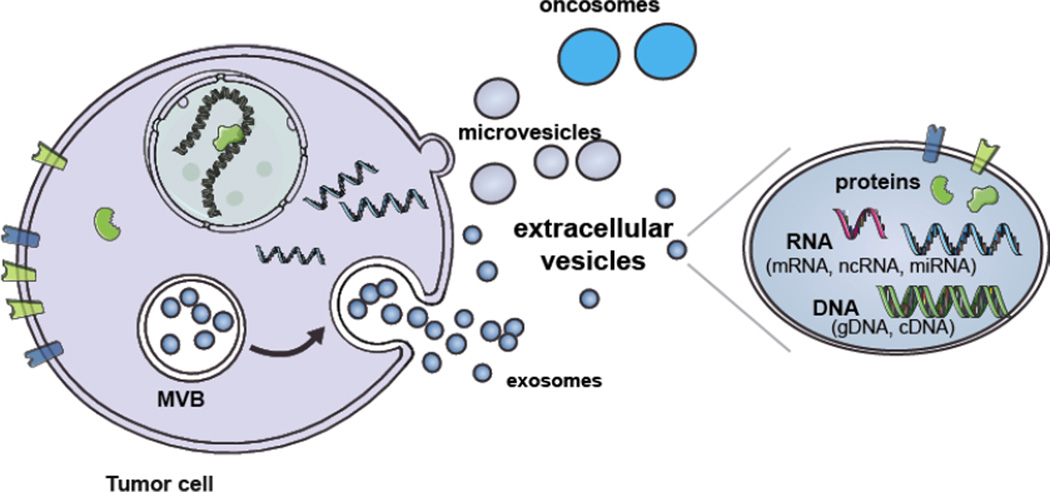
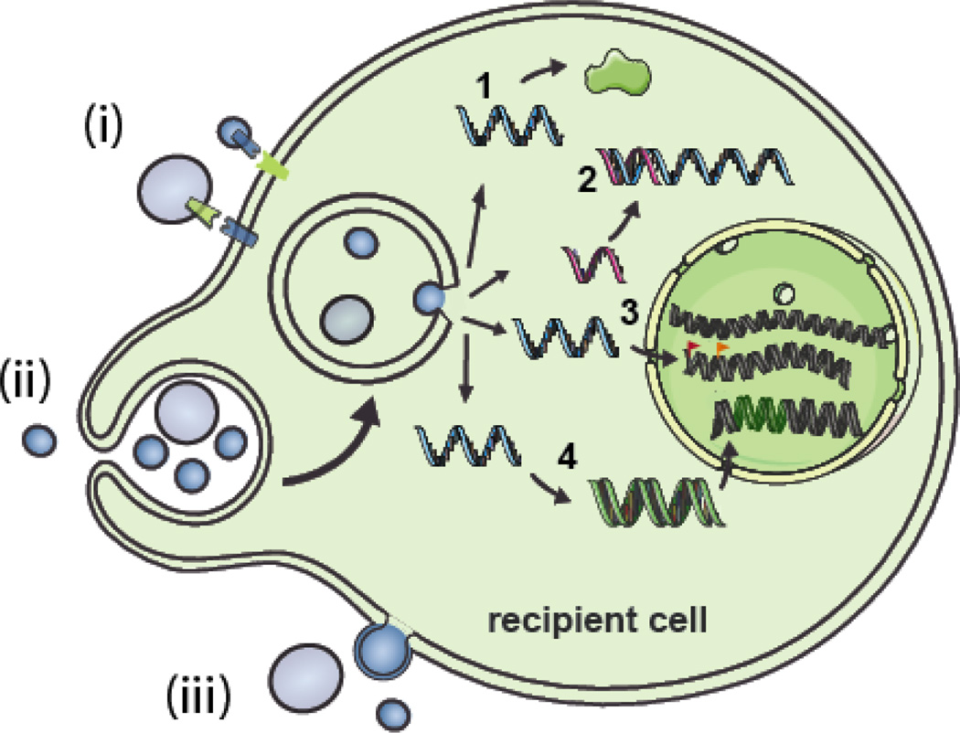
Different types of RNAs identified thus far represent a diverse group of macromolecules that are involved in the regulation of different biological processes. RNA is generally thought to be localized primarily in the nucleus and cytoplasm; however, some types of RNA have been detected in the extracellular milieu. These extracellular RNA (exRNA) molecules are protected from degradation and it is now widely accepted that extracellular vesicles and ribonucleoprotein particles serve as transport vehicles for exRNA among cells. The functional consequence of this transfer of genetic information probably encompasses a broad range of normal developmental and physiologic processes in many organisms. This review will focus on the role of exRNA communication in cancer. We will focus on different types of RNA species identified and characterized within tumor-derived extracellular vesicles. Further, we will describe the role of exRNAs in cancer progression, as well as their potential for use as diagnostic biomarkers and therapeutic tools for monitoring and treating cancer, respectively.

Keywords: Cancer; Extracellular vesicles; RNA.


Conflict of interest statement

Conflict of interest disclosure

The authors declare that there are no conflicts of interest.

Figures

Figure 1
Figure 1. Biogenesis and cargo of EVs
Extracellular vesicles are comprised of several different types of vesicles including exosomes, microvesicles and oncosomes. Exosomes are formed by the internalization of the endocytic membrane and formation of MVB inside the cell. The fusion of the MVBs with the plasma membrane results in the release of exosomes into the extracellular milieu. Microvesicles are formed by the outward budding of the plasma membrane and are directly released into the extracellular milieu. Oncosomes are a larger type of EV released by cancer cells through budding of the plasma membrane. EVs contain a variety of proteins, RNA species and types of DNA. (Modified from [62].)
Figure 2
Figure 2. Interactions of EVs with cells and the potential functional roles of exRNA
EVs can interact with recipient cells in several ways. i) The EV membrane (bearing ligands) can come in contact with receptors on the cell membrane thereby activating signaling pathways. ii) The EV membrane can fuse with the cell membrane or iii) be taken up by endocytosis/pinocytosis/phagocytosis. Depending on the type of exRNA present in the EVs these RNA molecules can have multiple effects on the phenotype of the recipient cell. 1) mRNAs can lead to translation of new proteins. 2) miRNAs can inhibit translation by binding to their mRNA targets. 3) ncRNAs can potentially alter genome methylation patterns and histone modifications by re-targeting epigenetic modifiers leading to altered gene expression patterns. 4) Repetitive elements may be integrated into the cell genome after reverse transcription. (Modified from [62].)
Figure 3
Figure 3. Generation of therapeutic Evs
A) EVs can be isolated from cultured cells derived from a patient, e.g. dendritic cells, hematopoietic cells and fibroblasts, and then loaded with a therapeutic RNA, e.g. siRNA or miRNA ex vivo, thus providing a non-immunogenic vehicle for RNA delivery. The endogenous contents of these EVs will vary depending on the cell type from which they are derived and in some cases these contents may also promote therapy. B) Cells can also be transfected in culture with an expression cassette for an mRNA encoding a targeting ligand (1) or therapeutic protein for incorporation into the EVs. Other RNAs, e.g. shRNA, miRNA or mRNAs can be expressed at high levels in the donor cell, possibly including EV incorporation signals, and used to deliver therapeutic RNAs.

References

    1. Kolodny GM. Evidence for transfer of macromolecular RNA between mammalian cells in culture. Exp Cell Res. 1971;65:313–324. - PubMed
    1. Valadi H, Ekström K, Bossios A, Sjöstrand M, Lee JJ, Lötvall JO. Exosome-mediated transfer of mRNAs and microRNAs is a novel mechanism of genetic exchange between cells. Nat Cell Biol. 2007;9:654–659. - PubMed
    1. Skog J, Würdinger T, van Rijn S, Meijer D, Gainche L, Curry WTJ, et al. Glioblastoma microvesicles transport RNA and protein that promote tumor growth and provide diagnostic biomarkers. Nat Cell Biol. 2008;10:1470–1476. - PMC - PubMed
    1. Baj-Krzyworzeka M, Szatanek R, Weglarczyk K, Baran J, Urbanowicz B, Branski P, et al. Tumour-derived microvesicles carry several surface determinants and mRNA of tumour cells and transfer some of these determinants to monocytes. Cancer Immunol Immunother. 2006;55:808–818. - PMC - PubMed
    1. Panagopoulos K, Cross-Knorr S, Dillard C, Pantazatos D, Del Tatto M, Mills D, et al. Reversal of chemosensitivity and induction of cell malignancy of a non-malignant prostate cancer cell line upon extracellular vesicle exposure. Mol Cancer. 2013;12:118. - PMC - PubMed

Publication types