

Macroscopic and histological variations in the cellular tapetum in dogs
- PMID: 24784162
- PMCID: PMC4155189
- DOI: 10.1292/jvms.14-0132
Macroscopic and histological variations in the cellular tapetum in dogs
Abstract
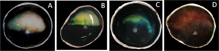
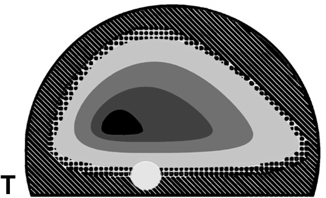
We aimed to document macroscopic variations in the cellular tapetum in the dog, to provide a histologic description of the macroscopic results and to evaluate the correlation between the macroscopic appearance and aging. Fifty three dogs including 5 beagles, 1 Chihuahua and 47 mixed breeds of each gender were used. For a macroscopic study, the fresh tapetal fundi were photographed using digital camera. For a histological study, the glutaraldehyde-formalin fixed eyes were embedded in nitrocellulose and stained with hematoxylin-eosin or thionine. The normal tapetum was triangular with the rounded angles and the smooth contour. The atypical tapetum was smaller and more variable in shape, contour and color than the normal one. In severe cases, the fundus was devoid of the tapetum. The atypical tapetum tended to increase in frequency with aging. Retinal pigment epithelial cells on the normal tapetum were unpigmented. In the eye with the atypical tapetum, regardless of tapetal size and shape, unpigmented retinal pigment epithelial cells showed a similar distribution to that on the normal tapetum, even in a dog without a tapetum. Although there is a congenitally hypoplastic tapetum, the atypical tapetum tends to increase in incidence and severity with aging.
Figures


Similar articles
-
Relationship between distribution of tapetum fibrosum and retinal pigment epithelium in the sheep eye.J Vet Med Sci. 2010 Feb;72(2):211-5. doi: 10.1292/jvms.09-0413. Epub 2009 Nov 27. J Vet Med Sci. 2010. PMID: 19942810
-
The fibrous tapetum of the horse eye.J Anat. 2013 Nov;223(5):509-18. doi: 10.1111/joa.12100. Epub 2013 Sep 15. J Anat. 2013. PMID: 24102505 Free PMC article.
-
Tapetal dysplasia in a Swedish Vallhund dog.Vet Ophthalmol. 2013 Jul;16 Suppl 1:145-50. doi: 10.1111/vop.12031. Epub 2013 Feb 13. Vet Ophthalmol. 2013. PMID: 23406395
-
Grouped retinae and tapetal cups in some Teleostian fish: occurrence, structure, and function.Prog Retin Eye Res. 2014 Jan;38:43-69. doi: 10.1016/j.preteyeres.2013.10.001. Epub 2013 Oct 22. Prog Retin Eye Res. 2014. PMID: 24157316 Review.
-
Review on tapetal ultrastructure in angiosperms.Planta. 2023 Apr 21;257(6):100. doi: 10.1007/s00425-023-04138-8. Planta. 2023. PMID: 37084157 Review.
Cited by
-
Abnormal Appearance of the Area Centralis in Labrador Retrievers With an ABCA4 Loss-of-function Mutation.Transl Vis Sci Technol. 2022 Feb 1;11(2):36. doi: 10.1167/tvst.11.2.36. Transl Vis Sci Technol. 2022. PMID: 35201338 Free PMC article.
-
Morphology and Histology of the Orbital Region and Eye of the Asiatic Black Bear (Ursus thibetanus)-Similarities and Differences within the Caniformia Suborder.Animals (Basel). 2022 Mar 22;12(7):801. doi: 10.3390/ani12070801. Animals (Basel). 2022. PMID: 35405790 Free PMC article.
-
Dog eye movements are slower than human eye movements.J Eye Mov Res. 2020 Feb 5;12(8):10.16910/jemr.12.8.4. doi: 10.16910/jemr.12.8.4. J Eye Mov Res. 2020. PMID: 33828775 Free PMC article.
-
Morphological and Morphometric Analysis of Canine Choroidal Layers Using Spectral Domain Optical Coherence Tomography.Int J Environ Res Public Health. 2023 Feb 10;20(4):3121. doi: 10.3390/ijerph20043121. Int J Environ Res Public Health. 2023. PMID: 36833819 Free PMC article.
-
Anatomical and morphometric evaluation of the orbit, eye tunics, eyelids and orbital glands of the captive females of the South African painted dog (Lycaon pictus pictus Temminck, 1820) (Caniformia: Canidae).PLoS One. 2021 Apr 19;16(4):e0249368. doi: 10.1371/journal.pone.0249368. eCollection 2021. PLoS One. 2021. PMID: 33872321 Free PMC article.
References
-
- Bellhorn R. W., Bellhorn M. B., Swarm R. L., Impellizzeri C. W.1975. Hereditary tapetal abnormality in the beagle. Ophthalmol. Res. 7: 250–260. doi: 10.1159/000264758 - DOI
-
- Diesem C.1975. Carnivore sense organs and common integument. pp. 1741–1768. In: Sisson and Grossman’s The Anatomy of Domestic Animals, 5th ed. (Getty, R. ed.), WB Saunders, Philadelphia.
MeSH terms
LinkOut - more resources
Full Text Sources
Other Literature Sources
Medical