

Improvement of bone regeneration capability of ceramic scaffolds by accelerated release of their calcium ions
- PMID: 24784792
- PMCID: PMC4229868
- DOI: 10.1089/ten.TEA.2012.0726
Improvement of bone regeneration capability of ceramic scaffolds by accelerated release of their calcium ions
Abstract
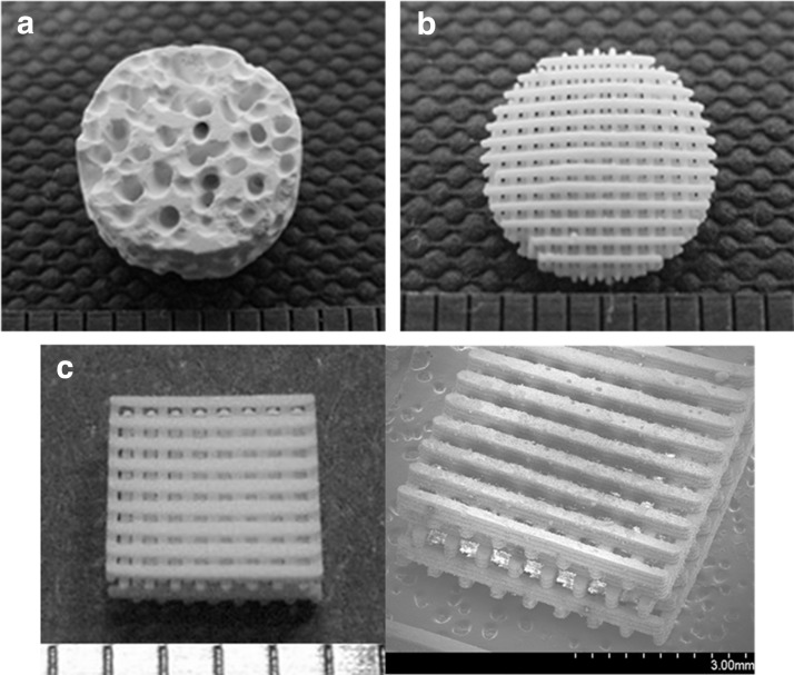
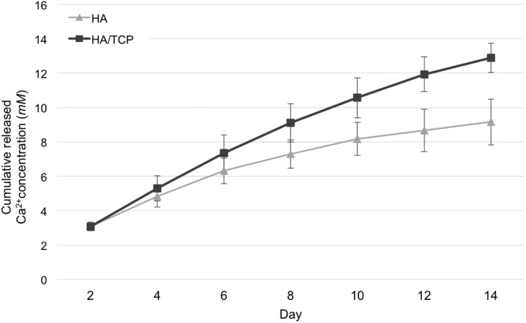
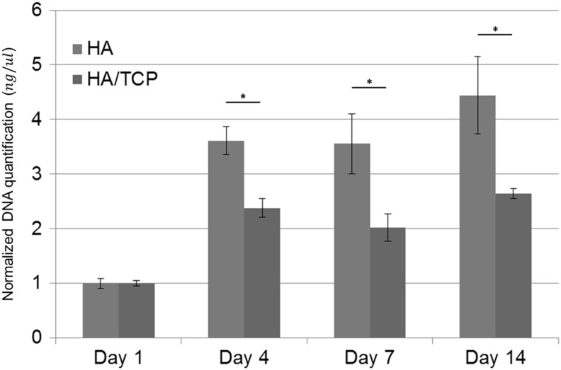
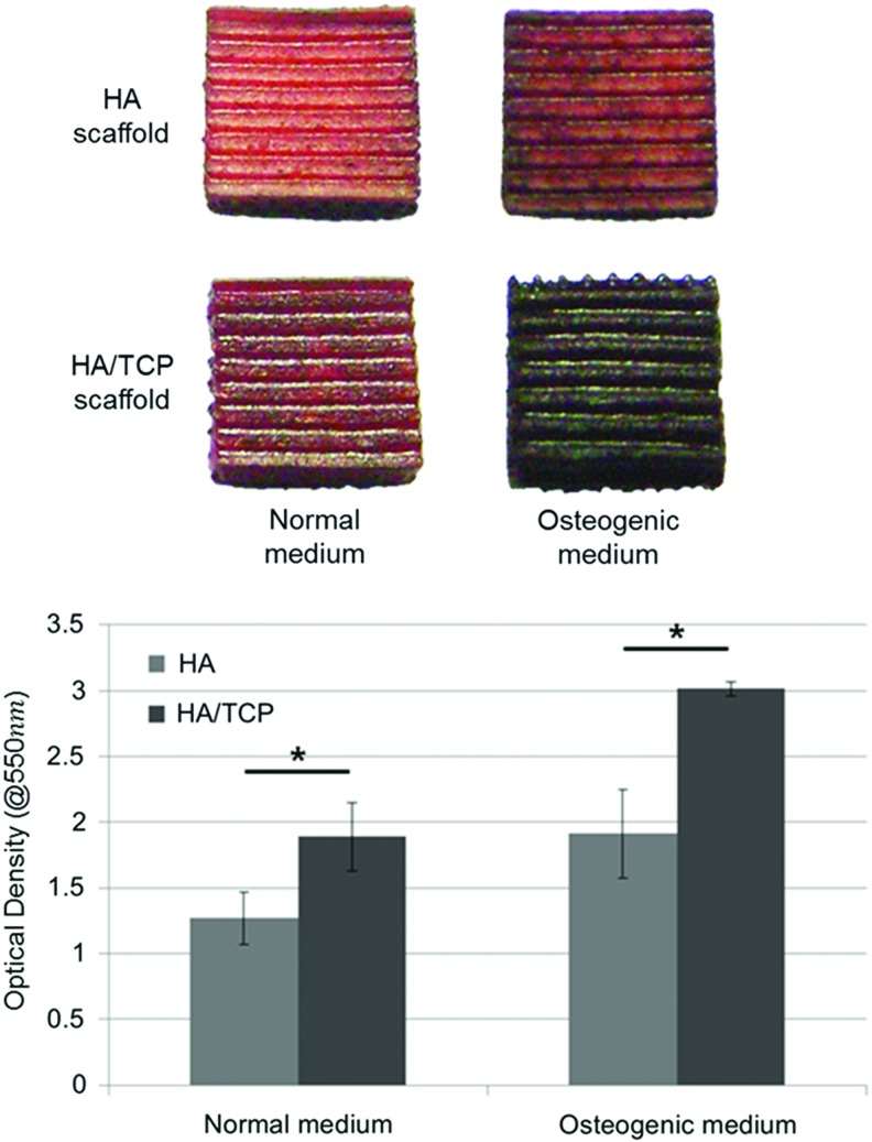
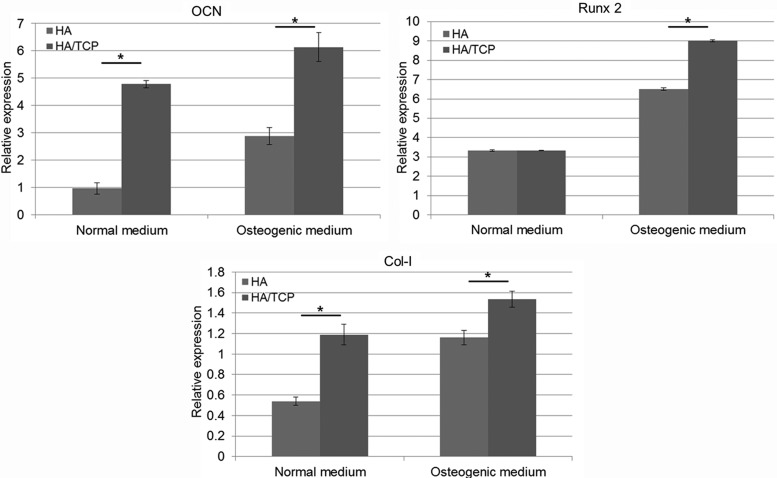
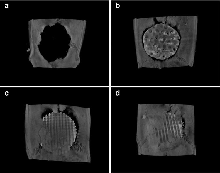
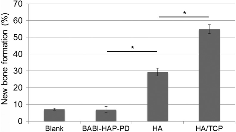
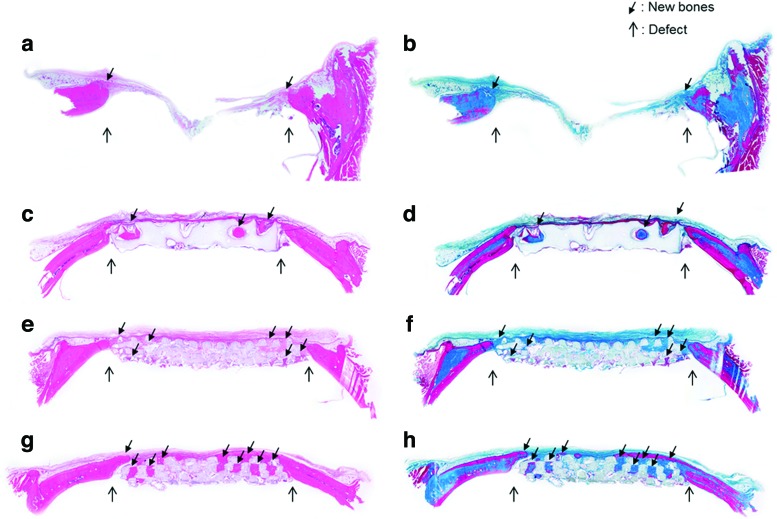
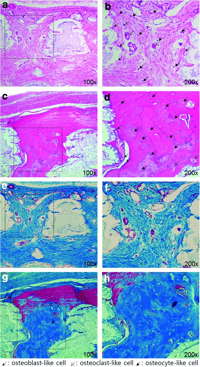
To regenerate the bone tissue, the fabrication of scaffolds for better tissue regeneration has attracted a great deal of attention. In fact, growth factors are already used in clinical practice and are being investigated for enhancing the capacity for bone tissue regeneration. However, despite their strong osteoinductive activity, these growth factors have several limitations: safety issues, high treatment costs, and the potential for ectopic bone formation. The aim of this study was therefore to develop ceramic scaffolds that could promote the capacity for bone regeneration without growth factors. Three-dimensional ceramic scaffolds were successfully fabricated from hydroxyapatite (HA) and tricalcium phosphate (TCP) using projection-based microstereolithography, which is an additive manufacturing technology. The effects of calcium ions released from ceramic scaffolds on osteogenic differentiation and bone regeneration were evaluated in vitro and in vivo. The osteogenesis-related gene expression and area of new bone formation in the HA/TCP scaffolds was higher than those in the HA scaffolds. Moreover, regenerated bone tissue in HA/TCP scaffolds were more matured than that in HA scaffolds. Through this study, we were able to enhance the bone regeneration capacity of scaffolds not by growth factors but by calcium ions released from the scaffolds. Ceramic scaffolds developed in this study might be useful for enhancing the capacity for regeneration in complex bone defects.
Figures
References
-
- Einhorn T.A.The cell and molecular biology of fracture healing. Clin Orthop Relat Res 355Suppl,S7, 1998 - PubMed
-
- Petite H., Viateau V., Bensaid W., Meunier A., de Pollak C., Bourguignon M., et al. Tissue-engineered bone regeneration. Nat Biotechnol 18,959, 2000 - PubMed
-
- Damien C.J., and Parsons J.R.Bone graft and bone graft substitutes: a review of current technology and applications. J Appl Biomater 2,187, 1991 - PubMed
-
- Hollister S.J.Porous scaffold design for tissue engineering. Nat Mater 4,518, 2005 - PubMed
-
- Langer R., and Vacanti J.P.Tissue engineering. Science 260,920, 1993 - PubMed
Publication types
MeSH terms
Substances
LinkOut - more resources
Full Text Sources
Other Literature Sources
Medical
Molecular Biology Databases
