

Hounsfield Units: a new indicator showing maxillary resistance in rapid maxillary expansion cases?
- PMID: 24784845
- PMCID: PMC8634823
- DOI: 10.2319/111013-823.1
Hounsfield Units: a new indicator showing maxillary resistance in rapid maxillary expansion cases?
Abstract
Objective: To determine if density measurements of several maxillary regions in Hounsfield Units (HU) and outcomes of rapid maxillary expansion (RME) are correlated. Is correlation powerful enough to give us direct information about maxillary resistance to RME?
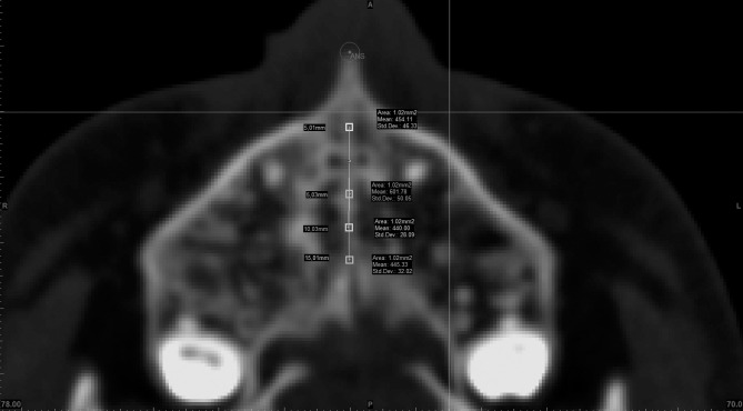
Materials and methods: Twenty-two computed tomographic (CT) scans (14 years) are used in this archive study. Two CT records were collected, one before RME (T1) and one after 3 months of retention period (T2). Maxillary measurements were made using dental and skeletal landmarks in first molar and first premolar slides to measure the effects of RME. Density of midpalatal suture (MPSD) and segments of maxillary bone is measured in HU at T1. Correlation analysis was conducted between density measurements and maxillary variables. Regression analysis was then performed for variables that showed positive correlation.
Results: There was no correlation between density and skeletal measurements. Intermolar angle (ImA) in molar slice showed statistically significant correlation with density measurements. The ImA variable showed the highest correlation with MPSD in frontal section (r = 0.669, P < .01).
Conclusions: There is correlation of 32.1-43.3% between density measurements and ImA increase. Our density measurements explain a certain percentage of ImA increase, but density is not the only and definitive indicator of changes after RME.
Keywords: Hounsfield Units; Rapid maxillary expansion; Suture density.
Figures


References
-
- Timms DJ. Rapid Maxillary Expansion. Chicago, Ill: Quintessence Publishing; 1981. pp. 91–94.
-
- Litsas G, Ari-Demirkaya A. Growth indicators in orthodontic patients. Part 2: comparison of cervical bone age to hand-wrist skeletal age. Relationship with chronological age. Eur J Paediatr Dent. 2010;11:176–180. - PubMed
-
- Wehrbein H, Yildizhan F. The mid-palatal suture in young adults. A radiological-histological investigation. Eur J Orthod. 2001;23:105–114. - PubMed
-
- Walker L, Enciso R, Mah J. Three-dimensional localization of maxillary canines with cone-beam computed tomography. Am J Orthod Dentofacial Orthop. 2005;128:418–423. - PubMed
Publication types
MeSH terms
LinkOut - more resources
Full Text Sources
Medical
