. 2014 May 1;10(5):e1004276.
doi: 10.1371/journal.pgen.1004276. eCollection 2014 May.

Revertant mutation releases confined lethal mutation, opening Pandora's box: a novel genetic pathogenesis

Affiliations

Revertant mutation releases confined lethal mutation, opening Pandora's box: a novel genetic pathogenesis

Yasushi Ogawa et al. PLoS Genet. .

Abstract

When two mutations, one dominant pathogenic and the other "confining" nonsense, coexist in the same allele, theoretically, reversion of the latter may elicit a disease, like the opening of Pandora's box. However, cases of this hypothetical pathogenic mechanism have never been reported. We describe a lethal form of keratitis-ichthyosis-deafness (KID) syndrome caused by the reversion of the GJB2 nonsense mutation p.Tyr136X that would otherwise have confined the effect of another dominant lethal mutation, p.Gly45Glu, in the same allele. The patient's mother had the identical misssense mutation which was confined by the nonsense mutation. The biological relationship between the parents and the child was confirmed by genotyping of 15 short tandem repeat loci. Haplotype analysis using 40 SNPs spanning the >39 kbp region surrounding the GJB2 gene and an extended SNP microarray analysis spanning 83,483 SNPs throughout chromosome 13 in the family showed that an allelic recombination event involving the maternal allele carrying the mutations generated the pathogenic allele unique to the patient, although the possibility of coincidental accumulation of spontaneous point mutations cannot be completely excluded. Previous reports and our mutation screening support that p.Gly45Glu is in complete linkage disequilibrium with p.Tyr136X in the Japanese population. Estimated from statisitics in the literature, there may be approximately 11,000 p.Gly45Glu carriers in the Japanese population who have this second-site confining mutation, which acts as natural genetic protection from the lethal disease. The reversion-triggered onset of the disesase shown in this study is a previously unreported genetic pathogenesis based on Mendelian inheritance.


Conflict of interest statement

The authors have declared that no competing interests exist.

Figures

Figure 1
Figure 1. Sequence and haplotype analysis of the present case of KID syndrome.
(A) Clinical manifestations of the patient are shown. Marked hyperkeratosis of the palms and soles is seen. (B) Identification of c.134G>A and c.408C>A mutations in the patient and her parents. The patient is compound heterozygous for the two mutations. (C) Haplotype analysis of the family members. Fourteen heterozygous SNPs spanning >39 kbp surrounding the GJB2 gene are identified and assembled into three contigs (shown in parenthesis). The nucleotides altered by the c.134G>A and c.408C>A mutations are boxed. The altered nucleotides are in red. The patient and her parents share a single haplotype (top; blue background). The patient has a unique haplotype (bottom; yellow background) that is not harbored by either parent. The maternally unique haplotype is shown in orange.
Figure 2
Figure 2. Configurations and topological mapping of the GJB2 mutations in the family.
(A) Structures of Cx26 mutants. The mutations/variants found in each allele of the family members are shown. p.Val27Ile and p.Glu114Gly are non-pathological variants. (B) Topological mapping of the Cx26 mutations. p.Gly45Glu (red) is located in the first extracellular loop domain and is thought to affect the channel activity of gap junctions. TM1–4: transmembrane domain 1–4; E1–2: extracellular domain 1–2; NT: N terminus; CT: C terminus.
Figure 3
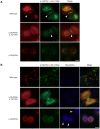
Figure 3. The p.Tyr136X mutation confines the effect of the pGly45Glu mutation.
(A) Gap junction formation by the transfected Cx26 variants. Each panel contains two co-transfected cells connected to each other. Wild-type, p.Gly45Glu/p.Tyr136X and p.Gly45Glu mutants of Cx26 were tagged with monomeric Red Fluorescent Protein (mRFP) and co-transfected with Green Fluorescent Protein (EGFP)-tagged Cx26 p.Val27Ile/p.Glu114Gly into HeLa cells as indicated. Gap junction formation sites are indicated by arrowheads. The combination of WT Cx26 and Cx26 p.Val27Ile/p.Glu114Gly (top row) results in gap junctions that consist of both Cx26 proteins (yellow signal). The combination of Cx26 p.Gly45Glu/p.Tyr136X and Cx26 p.Val27Ile/p.Glu114Gly (middle row) results in gap junctions with only Cx26 p.Val27Ile/p.Glu114Gly (green signal). No apparent gap junction formation is seen when Cx26 p.Gly45Glu and Cx26 p.Val27Ile/p.Glu114Gly are cotransfected (bottom row). (B) Aberrant gate opening detected with neurobiotin uptake assay. Fluorescent-tagged Cx26s were cotransfected into HeLa cells as indicated, and treated with neurobiotin in a calcium-free condition. Uptake was detected with AlexaFluor350 streptoavidin dye (blue). Aberrant uptake of neurobiotin is observed only in cells cotransfected with Cx26 p.Gly45Glu and Cx26 p.Val27Ile/p.Glu114Gly (bottom row).
Figure 4
Figure 4. Schematic of the mechanism whereby the p.Tyr136X mutation confines the effect of the p.Gly45Glu mutation.
The truncated Cx26 peptides produced from the mutant p.Gly45Glu/p.Tyr136X are not incorporated into connexons or gap junctions (middle row), although Cx26 peptides derived from the mutant p.Gly45Glu are incorporated into connexons, resulting in aberrant gate opening and malformation of gap junctions (bottom row).

References

    1. Skinner BA, Greist MC, Norins AL (1981) The keratitis, ichthyosis, and deafness (KID) syndrome. Arch Dermatol 117: 285–289. - PubMed
    1. Richard G, Rouan F, Willoughby CE, Brown N, Chung P, et al. (2002) Missense mutations in GJB2 encoding connexin-26 cause the ectodermal dysplasia keratitis-ichthyosis-deafness syndrome. Am J Hum Genet 70: 1341–1348. - PMC - PubMed
    1. Janecke AR, Hennies HC, Gunther B, Gansl G, Smolle J, et al. (2005) GJB2 mutations in keratitis-ichthyosis-deafness syndrome including its fatal form. Am J Med Genet A 133A: 128–131. - PubMed
    1. Arita K, Akiyama M, Aizawa T, Umetsu Y, Segawa I, et al. (2006) A novel N14Y mutation in Connexin26 in keratitis-ichthyosis-deafness syndrome: analyses of altered gap junctional communication and molecular structure of N terminus of mutated Connexin26. Am J Pathol 169: 416–423. - PMC - PubMed
    1. Tsukada K, Nishio S, Usami S (2010) A large cohort study of GJB2 mutations in Japanese hearing loss patients. Clin Genet 78: 464–470. - PubMed

Publication types

LinkOut - more resources