. 2014 May 2;344(6183):527-32.
doi: 10.1126/science.1252651.

Chemical inhibition of NAT10 corrects defects of laminopathic cells

Affiliations

Chemical inhibition of NAT10 corrects defects of laminopathic cells

Delphine Larrieu et al. Science. .

Abstract

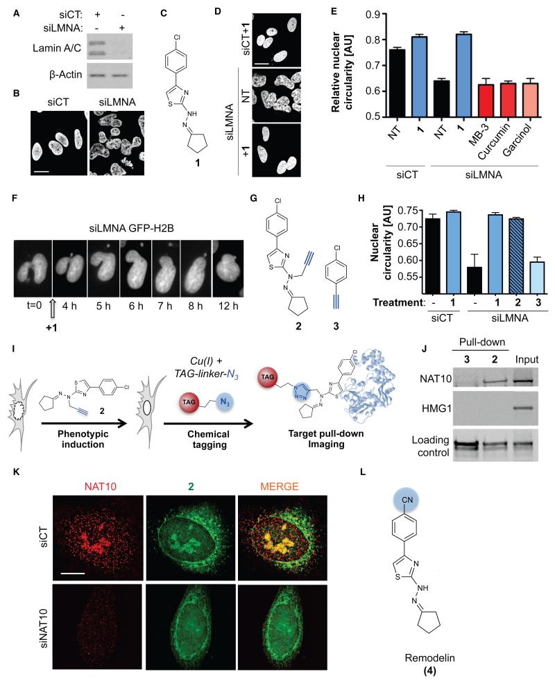
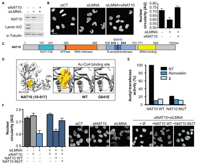
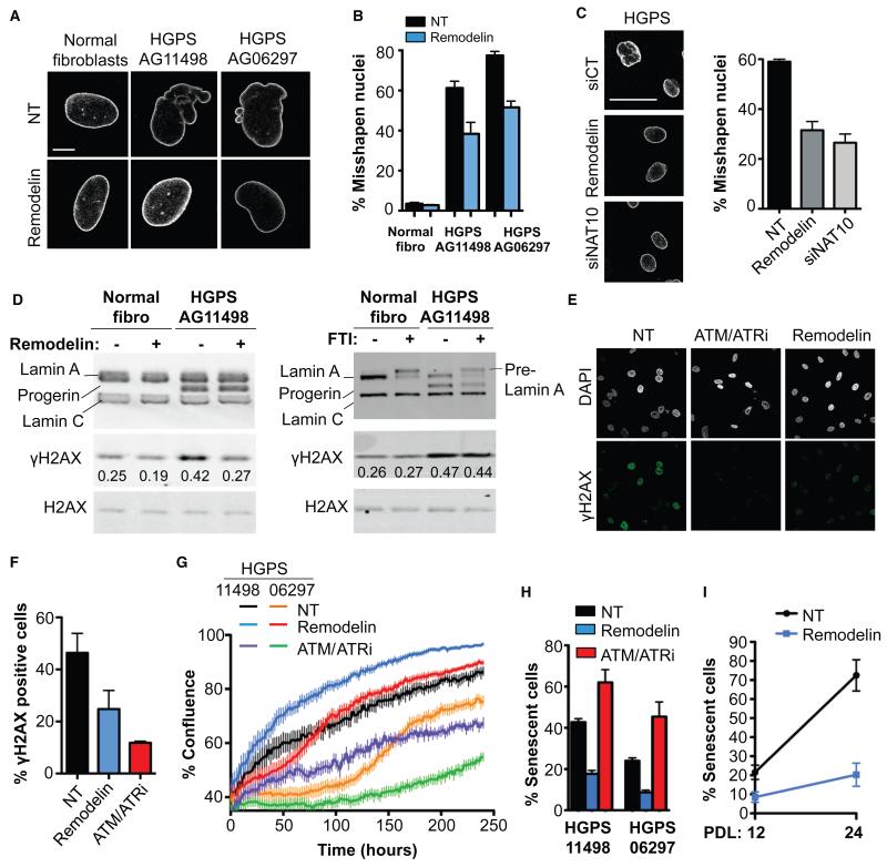
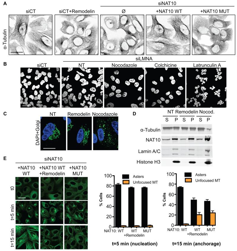
Down-regulation and mutations of the nuclear-architecture proteins lamin A and C cause misshapen nuclei and altered chromatin organization associated with cancer and laminopathies, including the premature-aging disease Hutchinson-Gilford progeria syndrome (HGPS). Here, we identified the small molecule "Remodelin" that improved nuclear architecture, chromatin organization, and fitness of both human lamin A/C-depleted cells and HGPS-derived patient cells and decreased markers of DNA damage in these cells. Using a combination of chemical, cellular, and genetic approaches, we identified the acetyl-transferase protein NAT10 as the target of Remodelin that mediated nuclear shape rescue in laminopathic cells via microtubule reorganization. These findings provide insights into how NAT10 affects nuclear architecture and suggest alternative strategies for treating laminopathies and aging.


Figures

Figure 1
Figure 1. A small molecule restores nuclear shape in Lamin A/C depleted cells and targets the acetyltransferase NAT10
A) Lamin A/C depletion (siLMNA) in U2OS cells compared to negative control (siCT). B) Nuclear shape observed by DAPI staining. C) Molecular structure of 4-(4-chlorophenyl)-2-(2-cyclopentylidenehydrazinyl)thiazole (1). D) Nuclear shape rescue observed by DAPI staining after treatment with 1. E) Quantification of nuclear circularity in non-treated (NT) cells or cells treated with the indicated compounds (means of three independent experiments with n>212 ± s.d.). F) Live imaging pictures of nuclear shape rescue in GFP-H2B expressing U2OS cells transfected with siLMNA and treated with 1. G) Molecular structure of clickable analogue 2 and clickable inactive control molecule 3. H) Quantification of U2OS nuclear circularity (means of three independent experiments with n>224 ± s.d.) I) Principle of click-chemistry strategy for small molecule tagging. J) Pull-down of clickable molecules 2 and 3 pre-incubated in U2OS cells and analysis of bound proteins. K) Representative high-resolution microscopy pictures of NAT10 (red) and fluorescently labeled 2 (green) in control or NAT10 depleted cells (siNAT10). Scale bars: 10 μm. L) Molecular structure of Remodelin (4), a stable and more potent analog of 1.
Figure 2
Figure 2. Inhibiting NAT10 activity by Remodelin mediates nuclear shape rescue of LMNA depleted cells
A) NAT10 and Lamin A/C depletion in U2OS cells. B) Nuclear shape visualized by DAPI staining (left) and quantification of nuclear circularity (right; means of three independent experiments with n>267 ± s.d.). Scale bar: 20 μm. C) Representation of NAT10 with its known domains. The G641E mutation identified in D) is indicated in dark blue and asterisked. D) Modelled 3D structure of human NAT10 residues 10-917 showing the acetyl-CoA binding site (left) and disruption of Ac-CoA binding by NAT10 G641E mutation (right) visualized with Swiss-Prot PDB Viewer. E) In vitro acetylation assay showing the activity of NAT10 towards tubulin. F) Quantification of nuclear circularity (left) in cells stably expressing siRNA-resistant FLAG-NAT10 WT or FLAG-NAT10 G641E (NAT10 MUT; means of three independent experiments with n>198 ± s.d.) and nuclear shape visualized by DAPI staining (right). Scale bar: 20 μm.
Figure 3
Figure 3. Remodelin targets NAT10 to improve nuclear shape and fitness of HGPS cells
A) Representative immunofluorescence (IF) pictures of Lamin A/C in HGPS cell lines compared to matched normal fibroblasts at the same population doubling. Scale bar: 10 μm. B) Quantification of misshapen nuclei upon Remodelin treatment (means of three independent experiments with n>213 ± s.e.m.). C) Lamin A/C staining in HGPS AG11498 cells (left) and quantification of misshapen nuclei (right; means of three independent experiments with n>176 ± s.e.m.). Scale bar: 50 μm. D) Western blotting analysis of γH2AX after Remodelin or FTI treatment. E) Immunofluorescence analysis of γH2AX staining upon Remodelin or ATM/ATR inhibition (ATM/ATRi). F) Quantification of γH2AX positive cells observed by IF (means of three independent experiments with n>127 ± s.e.m.). G) HGPS proliferation upon Remodelin or ATM/ATR-inhibitor treatment (means of nine replicates ± s.e.m). H) Quantification of senescence-associated β-galactosidase positive cells (means of three independent experiments with n>257 ± s.e.m.). I) Quantification of senescence-associated β-galactosidase positive cells in HGPS AG11498 after 8 days of Remodelin treatment at population doubling 12 (PDL 12) and after several weeks of Remodelin treatment and 12 cell divisions (PDL 24) (means of two independent experiments with n>298 ± s.d.).
Figure 4
Figure 4. Inhibiting NAT10 acetyltransferase activity modifies microtubule organisation to rescue nuclear shape defects
A) Microtubule network visualisation by inverted IF pictures of α-tubulin. B) Nuclear shape visualisation after treating cells with microtubule or actin cytoskeleton disrupting agents. C) Visualisation of nuclear shape (DAPI, blue) and Golgi (anti-Giantin, green) integrity in siLMNA cells. D) Fractionation of polymerized (P) and soluble (S) tubulin upon Remodelin or nocodazole (Nocod.) treatment. E) Microtubule regrowth in cells transfected with siNAT10 and expressing the indicated siRNA-resistant constructs. α-tubulin IF staining (left) shows nucleation phase: t=5 min and microtubule anchorage (t=15 min). Right: quantification of cells with indicated patterns (means of three independent experiments with n>103 ± s.e.m.).

References

    1. Worman HJ, Bonne G. “Laminopathies”: a wide spectrum of human diseases. Exp Cell Res. 2007 Jun 10;313:2121. - PMC - PubMed
    1. De Sandre-Giovannoli A, et al. Lamin a truncation in Hutchinson-Gilford progeria. Science. 2003 Jun 27;300:2055. - PubMed
    1. Eriksson M, et al. Recurrent de novo point mutations in lamin A cause Hutchinson-Gilford progeria syndrome. Nature. 2003 May 15;423:293. - PMC - PubMed
    1. Broers JL, et al. Nuclear A-type lamins are differentially expressed in human lung cancer subtypes. Am J Pathol. 1993 Jul;143:211. - PMC - PubMed
    1. Moss SF, et al. Decreased and aberrant nuclear lamin expression in gastrointestinal tract neoplasms. Gut. 1999 Nov;45:723. - PMC - PubMed

Publication types

MeSH terms