Review
. 2014 Jun;103(2):89-103.
doi: 10.1177/1457496914531927. Epub 2014 Apr 30.

Trauma-Induced Coagulopathy: An Institution's 35 Year Perspective on Practice and Research

Affiliations
Review

Trauma-Induced Coagulopathy: An Institution's 35 Year Perspective on Practice and Research

E Gonzalez et al. Scand J Surg. 2014 Jun.

Abstract

Introduction: Injury is the second leading cause of death worldwide, and as much as 40% of injury-related mortality is attributed to uncontrollable hemorrhage. This persists despite establishment of regionalized trauma systems and advances in the management of severely injured patients. Trauma-induced coagulopathy has been identified as the most common preventable cause of postinjury mortality.

Methods: A review of the current literature was performed by collecting PUBMED references related to trauma-induced coagulopathy. Data were then critically analyzed and summarized based on the authors' clinical and research perspective, as well as that reported by other institutions and researchers interested in trauma-induced coagulopathy. A particular focus was placed on those aspects of coagulopathy in which agreement among clinical and basic scientists is currently lacking; these include, pathophysiology, the role of blood components and factor therapy, and goal-directed assessment and management.

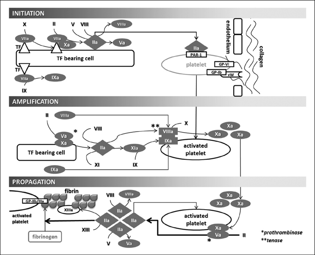
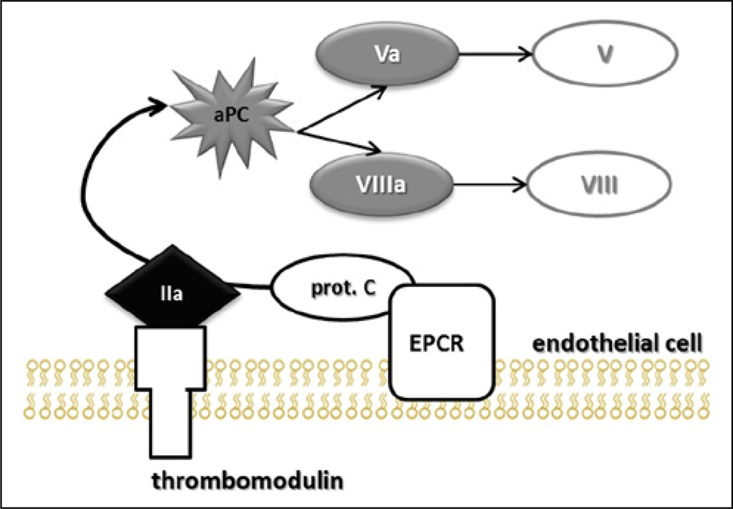
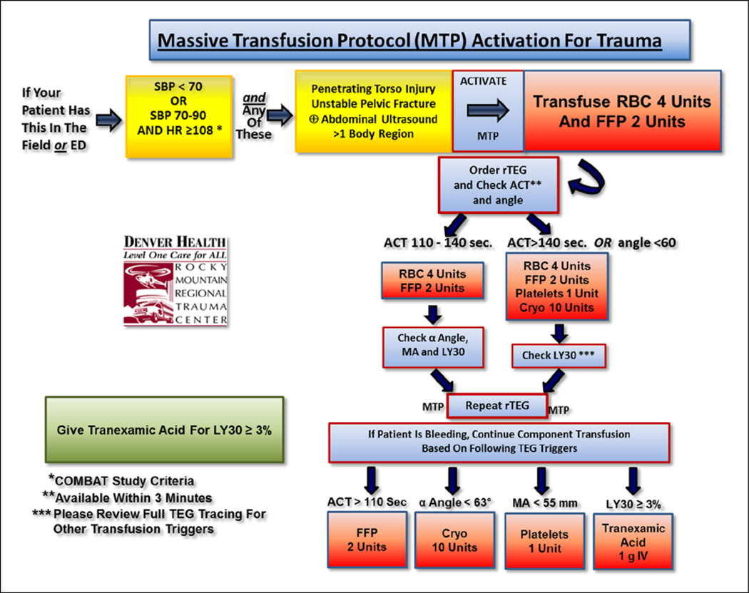
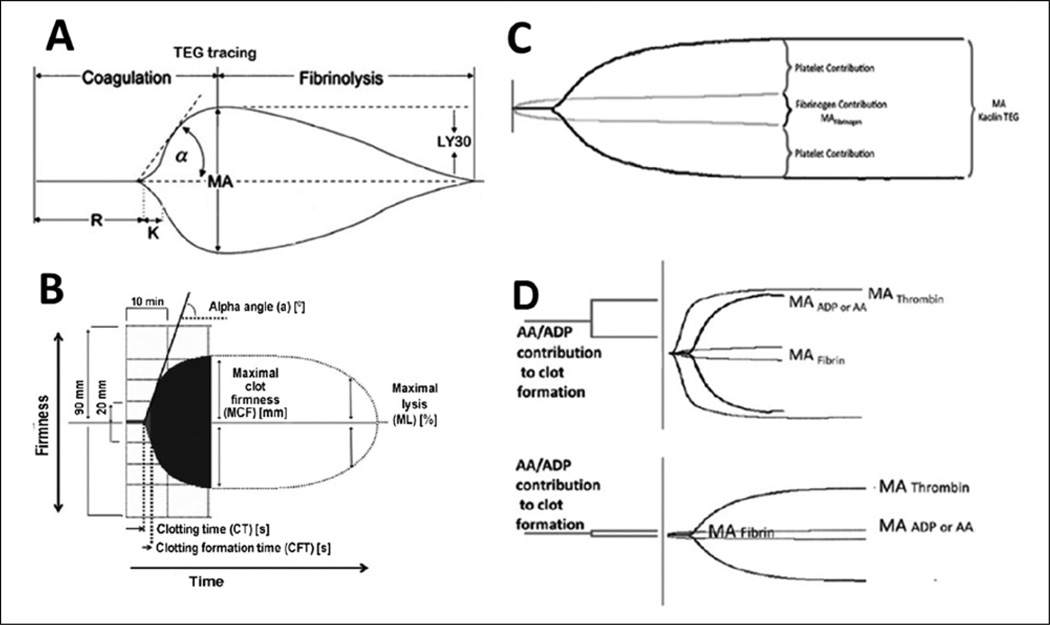
Results: Trauma-induced coagulopathy has been recognized in approximately one-third of trauma patients. There is a vast range of severity, and the emergence of viscoelastic assays, such as thrombelastography and rotational thromboelastogram, has refined its diagnosis and management, particularly through the establishment of goal-directed massive transfusion protocols. Despite advancements in the diagnosis and management of trauma-induced coagulopathy, much remains to be understood regarding its pathophysiology. The cell-based model of hemostasis has allowed for characterization of endothelial dysfunction, impaired thrombin generation, platelet dysfunction, fibrinolysis, endogenous anticoagulants such as protein-C, and antifibrinolytic proteins. These concepts collectively compose the contemporary, but still partial, understanding of trauma-induced coagulopathy.

Conclusion: Trauma-induced coagulopathy is a complex pathophysiological condition, of which some mechanisms have been characterized, but much remains to be understood in order to translate this knowledge into improved outcomes for the injured patient.

Keywords: Coagulopathy; ROTEM; fibrinolysis; hemorrhage; thrombelastography; transfusion; trauma.


Conflict of interest statement

DECLARATION OF CONFLICTING INTERESTS None.

Figures

Fig. 1
Fig. 1
This figure represents our current understanding of trauma-induced coagulopathy. On the left-hand column are those intrinsic and acquired factors that further potentiate TIC, including acidosis and hypothermia which were initially described with coagulopathy as the “bloody vicious cycle.” Hypoxia derived from hemorrhagic shock and tissue injury appears to be synergistic in driving TIC and activation of protein-C. DAMPS: damage-associated molecular pattern molecules; sCD40L: CD40 soluble platelet ligand; aPC: activated protein-C; tPA: tissue plasminogen activator; PAI: plasminogen activator inhibitor; TLR: toll-like receptor.
Fig. 2
Fig. 2
Cellular-based model of hemostasis. Initiation is triggered by TF bearing cells that activate factor VII. The TF/VIIa complex activates Xa and generates thrombin. During the initiation phase, thrombin activates factor V (which then associates with Xa to from the prothrombinase (Va/Xa) complex), factor VIII (which associates with IXa to form the tenase (VIIIa/IXa) complex during the amplification phase), and activates platelets via the protease-activated receptor (PAR-1). Simultaneously, platelets are being localized to the site of injury and activated by collagen via the platelet receptors GP Ib-IX-V (GPIb) and GP-VI. Factor IXa is also generated by TF/VIIa during the initiation phase, which can later directly participate in the propagation phase as part of the tenase complex, without requiring activation by XIa; this represents an overlap between the classically described mutually exclusive extrinsic and intrinsic pathways. During the amplification phase, the prothrombinase complex then generates thrombin that yields VIIIa and IXa, which form the tenase complex (VIIIa/IXa) on the activated platelet. The tenase complex will generate enough Xa to maintain pro-thombinase-mediated thrombin generation during the propagation phase. The amount of thrombin generated during the propagation phase is enough to cleave fibrinogen into fibrin, and also further generates Va and VIIIa (which further feeds prothrombinase and tenase), as well as XIIIa (which cross-links soluble fibrin into a stable clot). TF: tissue factor; PAR: protease-activated receptor; GP: glycoprotein.
Fig. 3
Fig. 3
Pathway for protein-C activation. Thrombomodulin is a transmembrane endothelial protein that binds thrombin. The thrombomodulin/thrombin complex is then responsible for protein-C activation (aPC). The endothelial protein-C receptor (EPCR) binds circulating protein-C to the endothelial surface, making it more susceptible to activation by the thrombomodulin/thrombin complex. aPC cleaves activated factors Va and VIIIa into their inactive forms V and VIII. aPC: activated protein-C; prot. C: protein-C; EPCR: endothelial protein-C receptor.
Fig. 4
Fig. 4
Mediators and inhibitors of fibrinolysis. In circulation, plasminogen adopts a closed, activation-resistant conformation. Upon binding to fibrin, plasminogen adopts an open form that can be converted into active plasmin by tissue plasminogen activator (tPA). Plasmin achieves fibrinolysis by cleaving fibrin into fibrin dimer products (FDPs). FDPs are quantified by the D-dimer laboratory assay. tPA activity is inhibited by plasminogen activator inhibitor 1 (PAI-1) and most circulating tPA is bound to this inactivator. Thrombin activatable fibrinolysis inhibitor (TAFI) down regulates fibrinolysis by modifying fibrin with removal of C-terminal lysines, making fibrin less susceptible to plasmin cleavage and decreasing the rate of fibrinolysis. Antiplasmin (AP) forms a stoichiometric complex with soluble plasmin directly inhibiting its activity. PAI: plasminogen activator inhibitor; tPA: tissue plasminogen activator; AP: antiplasmin; FDP: fibrin dimer products; TAFI: thrombin activatable fibrinolysis inhibitor.
Fig. 5
Fig. 5
Algorithm of massive transfusion protocol incorporating goal-directed therapy. MTP: massive transfusion protocol; RBC: red blood cell; FFP: fresh frozen plasma; rTEG: RAPID thrombelastography; ACT: activated clotting time; LY : lysis; TEG: thrombelastography; MA: maximum amplitude.
Fig. 6
Fig. 6
Classic illustration of the (A) thrombelastogram and (B) ROTEM viscoelastic tracings. Parameters for these tracings are described in Table 1. (C) Functional fibrinogen tracing demonstrating fibrin and platelet contribution to clot strength. (D) TEG platelet mapping. The amplitude (mm), depicted on the y-axis, in response to platelet agonists maximum amplitude-adenosine-diphosphate (MA-ADP) or maximum amplitude-arachidonic acid (MA-AA) is compared with the maximum hemostatic activity (maximum amplitude (MA)-Thrombin) minus fibrin contribution (MA-Fibrin). The superior tracing represents a normal contribution of platelet’s to the clot, and the inferior tracing represents compromised platelet function. TEG: thrombelastography; MA: maximum amplitude; AA: arachidonic acid; ADP: amplitude-adenosine-diphosphate.

References

    1. The State of US Health, 1990–2010: Burden of diseases, injuries, and risk factors. JAMA. 2013;310:591–608. - PMC - PubMed
    1. Sauaia A, Moore FA, Moore EE, et al. Epidemiology of trauma deaths: A reassessment. J Trauma. 1995;38:185–193. - PubMed
    1. Stefanini M. Basic mechanisms of hemostasis. Bull N Y Acad Med. 1954;30:239–277. - PMC - PubMed
    1. Scott R, Jr, Crosby WH. Changes in the coagulation mechanism following wounding and resuscitation with stored blood; A study of battle casualties in Korea. Blood. 1954;9:609–621. - PubMed
    1. Simmons RL, Collins JA, Heisterkamp CA. Coagulation disorders in combat casualties. I. Acute changes after wounding. II. Effects of massive transfusion. 3. Post-resuscitative changes. Ann Surg. 1969;169:455–482. - PMC - PubMed

LinkOut - more resources