. 2015 Feb 7;36(6):353-68a.
doi: 10.1093/eurheartj/ehu180. Epub 2014 Apr 30.

Genome-wide profiling of the cardiac transcriptome after myocardial infarction identifies novel heart-specific long non-coding RNAs

Affiliations

Genome-wide profiling of the cardiac transcriptome after myocardial infarction identifies novel heart-specific long non-coding RNAs

Samir Ounzain et al. Eur Heart J. .

Abstract

Aim: Heart disease is recognized as a consequence of dysregulation of cardiac gene regulatory networks. Previously, unappreciated components of such networks are the long non-coding RNAs (lncRNAs). Their roles in the heart remain to be elucidated. Thus, this study aimed to systematically characterize the cardiac long non-coding transcriptome post-myocardial infarction and to elucidate their potential roles in cardiac homoeostasis.

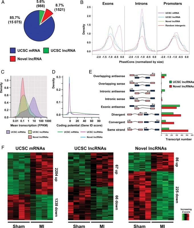
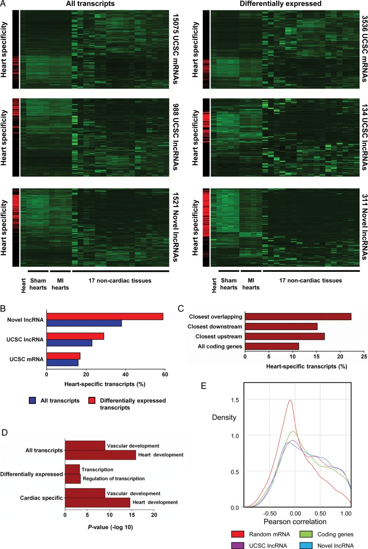
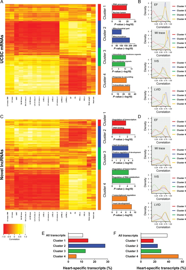
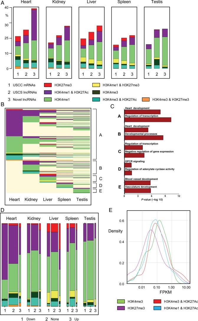
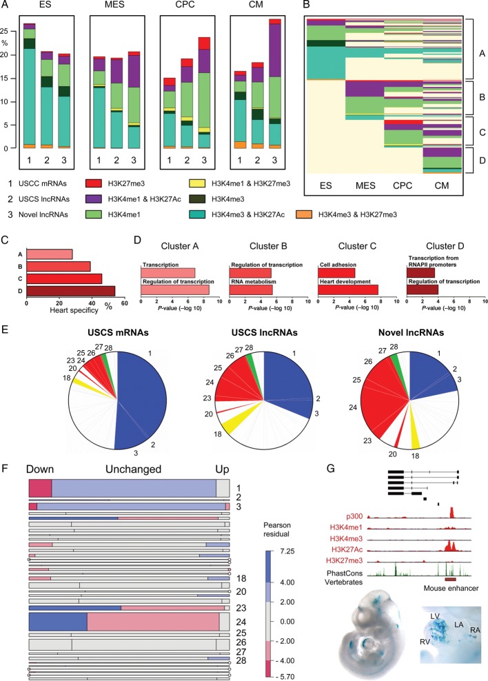
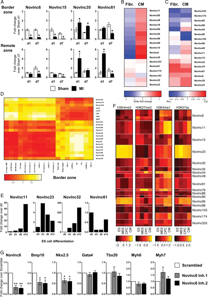
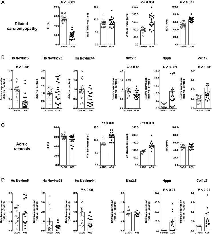
Methods and results: We annotated the mouse transcriptome after myocardial infarction via RNA sequencing and ab initio transcript reconstruction, and integrated genome-wide approaches to associate specific lncRNAs with developmental processes and physiological parameters. Expression of specific lncRNAs strongly correlated with defined parameters of cardiac dimensions and function. Using chromatin maps to infer lncRNA function, we identified many with potential roles in cardiogenesis and pathological remodelling. The vast majority was associated with active cardiac-specific enhancers. Importantly, oligonucleotide-mediated knockdown implicated novel lncRNAs in controlling expression of key regulatory proteins involved in cardiogenesis. Finally, we identified hundreds of human orthologues and demonstrate that particular candidates were differentially modulated in human heart disease.

Conclusion: These findings reveal hundreds of novel heart-specific lncRNAs with unique regulatory and functional characteristics relevant to maladaptive remodelling, cardiac function and possibly cardiac regeneration. This new class of molecules represents potential therapeutic targets for cardiac disease. Furthermore, their exquisite correlation with cardiac physiology renders them attractive candidate biomarkers to be used in the clinic.

Keywords: Heart failure; Long non-coding RNAs; Myocardial infarction; Next-generation sequencing; Transcriptome.


Figures

Figure 1
Figure 1
Global identification of long non-coding RNAs expressed in the heart during myocardial infarction . RNA-Seq during myocardial infarction was performed on border zone and sham operated samples to characterize the infarction associated transcriptome. (A) Pie chart showing composition of PolyA+ transcriptome, UCSC mRNAs (blue) UCSC long non-coding RNAs (green) and novel long non-coding RNAs (red). Transcript numbers in each group are indicated in brackets (B) Kernel density plots of phastCons score distribution of UCSC mRNA, UCSC long non-coding RNA, novel long non-coding RNA, and random intergenic sequence for exons, introns, and promoters. (C) Kernel density plot of the transcript abundance [fragments per kilobases per million reads (FPKM)] of UCSC mRNAs, UCSC long non-coding RNAs and novel long non-coding RNAs. (D) Kernel density plot of coding potential (Gene ID score). (E) Number of long non-coding RNA genes in orientations relative to nearby coding genes (F) Heatmaps showing hierarchical clustering of differentially expressed transcripts within the three RNA classes after myocardial infarction.
Figure 2
Figure 2
Tissue specificity of novel long non-coding RNAs. Novel long non-coding RNAs are more heart-specific and proximal to cardiac developmental genes. (A) Clustering of all and differentially expressed UCSC mRNAs, UCSC long non-coding RNAs and novel long non-coding RNAs across Sham-operated hearts, infarcted hearts, ENCODE heart and 17 non-cardiac mouse tissues. Left-hand panels highlight cardiac-specific transcripts in red. (B) Heart specificity of all and differentially expressed UCSC mRNAs, UCSC long non-coding RNAs and novel long non-coding RNAs. (C) Heart specificity of all UCSC coding genes compared with UCSC coding genes proximal to novel overlapping downstream and upstream long non-coding RNAs. (D) Enriched gene ontology terms for genes closest to all, differentially expressed and heart-specific novel long non-coding RNAs. (E) Kernel density plot displaying correlation of RNA expression for random gene pairs, neighbouring coding gene pairs, UCSC long non-coding RNA coding gene pairs and novel long non-coding RNA coding gene pairs during myocardial infarction.
Figure 3
Figure 3
Physiological clustering of the cardiac transcriptome. The cardiac transcriptome is highly correlated with cardiac physiological traits. (A) Clustering of UCSC mRNAs based on correlation of expression with echocardiography-derived physiological traits after myocardial infarction (Sham-operated and myocardial infarction samples were used for correlations). Clusters are illustrated right of the heatmap with top-enriched gene ontology terms for coding genes in each cluster displayed. (B) Kernel density plots of correlation of mRNA expression with selected physiological traits (ejection fraction, myocardial infarction trace, interventricular septal, and left ventricular internal diameter) within each cluster. (C) Clustering based on correlation of novel long non-coding RNA expression with echocardiography-derived physiological traits. Clusters are illustrated right of the heatmap with top-enriched terms for coding genes closest to novel long non-coding RNA. (D) Kernel density plots of correlation of long non-coding RNA expression with selected physiological traits (ejection fraction, myocardial infarction trace, interventricular septal, and left ventricular internal diameter) within each cluster. Heart specificity of UCSC mRNAs (E) and novel long non-coding RNAs (F) in clusters one to four.
Figure 4
Figure 4
Novel long non-coding RNAs are associated with specific chromatin states in the adult heart. Novel long non-coding RNAs are predominantly associated with adult heart-specific active enhancers (A) Bar charts of frequencies of UCSC mRNAs (1), UCSC long non-coding RNAs (2) and novel long non-coding RNAs (3) marked by H3K27me3, H3K4me3, H3K4me1, H3K4me3/H3K27Ac, H3K4me1/H3K27me3, H3K4me3/H3K27Ac, or H3K4me3/H3K27me3 in adult mouse heart, kidney, liver, spleen, and testis. (B) Epigenetic state map of all novel long non-coding RNAs marked by one of the above markers in at least one adult tissue including heart, kidney, liver, spleen, and testis. Rows are recursively clustered by marks (clusters A–E) in heart, kidney, liver, spleen, and testis. (C) Gene ontology terms significantly enriched in clusters A to E. (D) Mosaic plots showing the frequencies of transcripts, by differential expression category (vertical blocks) and by chromatin states (coloured cells within vertical blocks). Area of each coloured cell is proportional to frequency across all tissues; width of vertical bars is proportional to frequency of differential expression category; height of cell is proportional to frequency of chromatin state within expression category. (E) Enhancer-associated novel long non-coding RNAs (H3K4me1/H3K27Ac) tend to be more lowly expressed when compared with novel long non-coding RNAs with canonical promoter signatures (H3K4me3).
Figure 5
Figure 5
Inferring functions for novel long non-coding RNAs based on developmental chromatin state patterns. Novel long non-coding RNAs are more typically associated with chromatin state patterns linked to cardiac development and function. (A) Bar charts of frequencies of UCSC mRNAs (1), UCSC long non-coding RNAs (2) and novel long non-coding RNAs (3) marked by H3K27me3, H3K4me3, H3K4me1, H3K4me3/H3K27Ac, H3K4me1/H3K27me3, H3K4me3/H3K27Ac, and H3K4me3/H3K27me3 in embryonic stem cells, mesodermal precursors, cardiac precursor cells, and cardiomyocytes. (B) Epigenetic state map of all novel long non-coding RNA marked by one of the above markers in at least one lineage. Rows are recursively clustered by marks in embryonic stem, mesodermal precursors, cardiac precursor cell and cardiomyocyte (clusters A–D). (C) Heart specificity of chromatin-based clusters A–D. (D) Gene ontology terms significantly enriched in clusters A–D. (E) Pie charts illustrating associations of UCSC mRNAs, UCSC long non-coding RNAs and novel long non-coding RNAs with pre-determined chromatin state patterns according to Wamstad et al.. (F) Mosaic plot showing frequencies of transcripts by chromatin state cluster (horizontal blocks) and by differential expression status (cells within horizontal blocks). Area of each coloured cell is proportional to frequency across all lineages. Height of horizontal bar is proportional to frequency of cluster; width of cell is proportional to frequency of differential expression status within cluster. Cells are shaded according to the Pearson residual, providing a measure of enrichment within each cluster. Red shading indicates that the observed frequency is lower than expected while blue indicates greater than expected frequency. (G) The novlnc6 genomic locus is associated with a bonafide developmental enhancer (red box). Lower panel is a Lac-Z stained embryo and isolated heart with in vivo enhancer activity at E11.5. Stained embryo images were obtained from http://enhancer.lbl.gov/.
Figure 6
Figure 6
Selection and validation of novel long non-coding RNAs. High priority candidates were selected based on their association with unique and specific characteristics. See Supplementary material online, Table S5 (A) Quantitative RT–PCR analysis of selected novel long non-coding RNAs in Sham-operated (white bar) and infarcted heart (black bar), in the border and remote zones at 1 and 7 days post-infarction. (B) Heatmap representation of qRT–PCR analysis of long non-coding RNAs in cardiomyocytes and fibroblasts isolated from neonatal mouse hearts. (C) Heatmap representation of qRT–PCR analysis of long non-coding RNAs in the cytoplasmic or nuclear fractions of cardiomyocytes and fibroblasts from neonatal mouse hearts. Blue indicates nuclear enrichment and red cytoplasmic enrichment. (D) Heatmap representation of correlation between validated novel long non-coding RNA expression in the border zone and echocardiography-derived physiological traits after infarction. (E) Expression of selected novel long non-coding RNA in embryonic stem, mesodermal precursors, cardiac precursor cell and cardiomyocyte as measured by qRT–PCR. (F) Heatmaps of ChIP enrichment values of different chromatin modification at TSS region of validated novel long non-coding RNA in embryonic stem, mesodermal precursors, cardiac precursor cell, and cardiomyocyte. Colour scale is based on log10 of the ChIP signal. (G) Mouse neonatal cardiomyocytes were transfected with GapmeRs targeting a novlnc6 or random scrambled sequence. Cells were harvested 48 h post-transfection and assayed for Novlnc6, Bmp10, Nkx2–5, GATA4, Tbx20, Myh6, and Myh7 expression by qRT–PCR. Bars represent means ± SEM (n = 6 independent experiments). **P < 0.001; *P < 0.05.
Figure 7
Figure 7
Dysregulation of human long non-coding RNA orthologues in cardiac pathology. Human novel long non-coding RNA orthologues were detected and differentially modulated in pathological cardiac states. (A) Physiological parameters of the dilated cardiomyopathy patient cohorts. (B) Expression of Novlnc6, Novlnc23, Novlnc44, Nkx2.5, Nppa, and Col1a2 in heart samples from dilated cardiomyopathy patients. (C) Physiological parameters of the aortic stenosis patient cohorts. (D) Expression of Novlnc6, Novlnc23, Novlnc44, Nkx2.5, Nppa, and Col1a2 in heart samples from aortic stenosis patients.

References

    1. Roger VL, Go AS, Lloyd-Jones DM, Benjamin EJ, Berry JD, Borden WB, Bravata DM, Dai S, Ford ES, Fox CS, Fullerton HJ, Gillespie C, Hailpern SM, Heit JA, Howard VJ, Kissela BM, Kittner SJ, Lackland DT, Lichtman JH, Lisabeth LD, Makuc DM, Marcus GM, Marelli A, Matchar DB, Moy CS, Mozaffarian D, Mussolino ME, Nichol G, Paynter NP, Soliman EZ, Sorlie PD, Sotoodehnia N, Turan TN, Virani SS, Wong ND, Woo D, Turner MB American Heart Association Statistics C, Stroke Statistics S. Heart disease and stroke statistics--2012 update: a report from the American Heart Association. Circulation. 2012;125:e2–e220. - PMC - PubMed
    1. Hill JA, Olson EN. Cardiac plasticity. N Engl J Med. 2008;358:1370–1380. - PubMed
    1. van Berlo JH, Maillet M, Molkentin JD. Signaling effectors underlying pathologic growth and remodeling of the heart. J Clin Invest. 2013;123:37–45. - PMC - PubMed
    1. Lee TI, Young RA. Transcriptional regulation and its misregulation in disease. Cell. 2013;152:1237–1251. - PMC - PubMed
    1. Mercer TR, Mattick JS. Structure and function of long noncoding RNAs in epigenetic regulation. Nat Struct Mol Biol. 2013;20:300–307. - PubMed

Publication types