. 2014 Apr 30;6(234):234ra58.
doi: 10.1126/scitranslmed.3008085.

An acellular biologic scaffold promotes skeletal muscle formation in mice and humans with volumetric muscle loss

Affiliations

An acellular biologic scaffold promotes skeletal muscle formation in mice and humans with volumetric muscle loss

Brian M Sicari et al. Sci Transl Med. .

Abstract

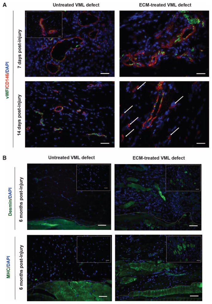
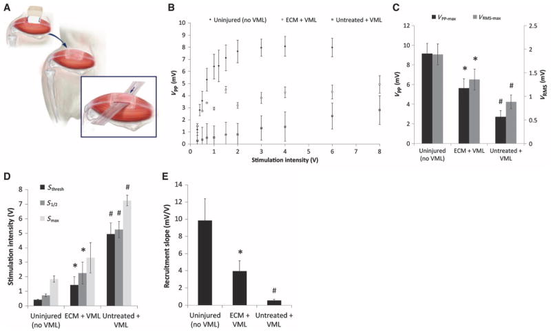
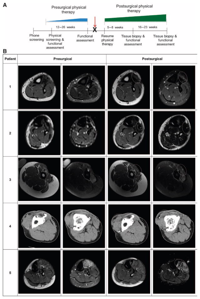
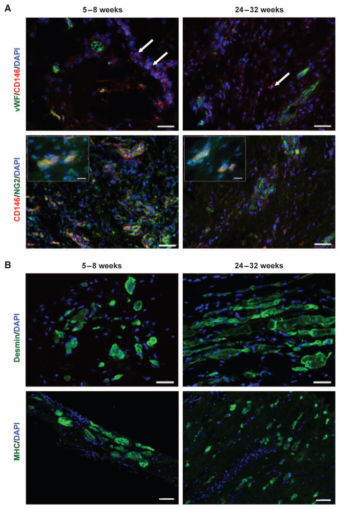
Biologic scaffolds composed of naturally occurring extracellular matrix (ECM) can provide a microenvironmental niche that alters the default healing response toward a constructive and functional outcome. The present study showed similarities in the remodeling characteristics of xenogeneic ECM scaffolds when used as a surgical treatment for volumetric muscle loss in both a preclinical rodent model and five male patients. Porcine urinary bladder ECM scaffold implantation was associated with perivascular stem cell mobilization and accumulation within the site of injury, and de novo formation of skeletal muscle cells. The ECM-mediated constructive remodeling was associated with stimulus-responsive skeletal muscle in rodents and functional improvement in three of the five human patients.


Conflict of interest statement

Competing interests: Since completion of the clinical study, S.F.B. has received honoraria from ACell Inc. for seminars. The other authors declare that they have no other competing interests.

Figures

Fig. 1
Fig. 1. Progenitor cells are present at the site of ECM scaffolds surgically placed within sites of mouse VML injury
(A) CD146+ PVSCs remained in their normal anatomic location, surrounding vascular structures (identified by vWF) at 7 days after injury in both untreated and ECM-treated defects in a mouse model of VML. Healthy, normal mouse skeletal tissue is shown in inset. After 14 days, PVSCs maintained their vascular association in untreated VML defects; conversely, in ECM-treated defects, CD146+ PVSCs were present outside their normal niche (arrows). (B) Untreated defects from the mouse model of VML showed no signs of skeletal muscle formation (desmin+, MHC+ cells), whereas ECM scaffold–treated defects showed desmin+, MHC+, and striated skeletal muscle cells at 6 months after injury. Skeletal muscle cells were identified along the interface with underlying native tissue (dotted white line) as well as throughout the center of the ECM implantation site (insets). Scale bars, 50 μm. DAPI, 4′,6-diamidino-2-phenylindole; MHC, major histocompatibility complex.
Fig. 2
Fig. 2. EMG at 6 months in healthy mouse muscle and in mice with untreated or ECM-treated VML defects
(A) Schematic representation of insulated EMG electrode placement within areas of remodeled tissue. (B) A range of stimulation intensities was applied to generate muscle recruitment curves from the evoked peak-to-peak voltage (VPP) response. (C) Maximal VPP and VRMS responses for each treatment. (D) Stimulation intensities required to evoke the minimum CMAP response (Sthresh), one-half the maximum response (S1/2), and the maximum response (Smax) for each treatment group. (E) Recruitment rate was calculated as the slope of the recruitment curve at S1/2. Data in (B) to (E) are means ± SEM (n = 8). *P < 0.05 versus untreated, #P < 0.05 versus healthy, Kruskal-Wallis non-parametric analysis of variance (ANOVA) with a post hoc Mann-Whitney U test and Sidak correction.
Fig. 3
Fig. 3. Study design timeline and imaging of affected extremity before and after surgical implantation in patients
(A) All patients completed a customized presurgical physical therapy program that focused specifically on their individual functional deficits and that was designed to maximize their individual potentials before surgical intervention (blue ramp progression, 12 to 26 weeks). The identification and maximization of functional parameters before surgery minimized the likelihood that any improvement after ECM intervention was solely the result of the physical therapy component of care. Patients then underwent the ECM scaffold surgical placement procedure (red arrow) and immediately returned to their presurgical physical therapy regimen (green ramp progression, 5 to 23 weeks). Ultrasound-guided biopsies were taken from the site of ECM scaffold implantation at 5 to 8 weeks for immunostaining. (B) Postsurgical images were taken at 6 months. Implantation site is denoted by dotted line. Patient 1: Pre- and postsurgical axial T1–weighted (left) and fat-saturated T2–weighted (right) MR images of the left distal calf. Patient 2: Presurgical axial T1–weighted (left) and short tau inversion recovery (STIR)–weighted (right); post-surgical axial T1–weighted (left) and T2-weighted (right) MR images of the distal calf. Patient 3: Pre-and postsurgical axial T1 (left) and fat-saturated T2 (right) MR images of the left mid-thigh. Patient 4: Pre- and postsurgical contiguous unenhanced axial CT imaging of the proximal (left) and distal (right) right thigh. Patient 5: Presurgical axial T1 (left) and fat-saturated T2 (right); postsurgical axial T1–weighted (left) and STIR-weighted (right) MR images of the proximal left calf.
Fig. 4
Fig. 4. Constructive tissue remodeling by ECM scaffolds in patients
Representative images are shown here, with images from each patient in fig. S2. (A) Biopsied human muscle tissue from ECM-treated VML defects at both 5 to 8 weeks and 24 to 32 weeks after scaffold implantation. PVSCs (CD146+NG2+) were present both within and outside of (arrows) their normal perivascular association (vWF+ regions). High-magnification insets show colocalization of CD146 and NG2 (inset scale bars, 10 μm). Scale bars, 50 μm. (B) Human muscle biopsies from the site of scaffold implantation at 5 to 8 weeks and 24 to 32 weeks after ECM scaffold implantation showed the formation of islands of desmin+ muscle cells. Scale bars, 50 μm.

References

    1. Huard J, Li Y, Fu FH. Muscle injuries and repair: Current trends in research. J Bone Joint Surg Am. 2002;84-A:822–832. - PubMed
    1. Yin H, Price F, Rudnicki MA. Satellite cells and the muscle stem cell niche. Physiol Rev. 2013;93:23–67. - PMC - PubMed
    1. Chargé SB, Rudnicki MA. Cellular and molecular regulation of muscle regeneration. Physiol Rev. 2004;84:209–238. - PubMed
    1. Järvinen T, Järvinen TL, Kääriäinen M, Kalimo H, Järvinen M. Muscle injuries: Biology and treatment. Am J Sports Med. 2005;33:745–764. - PubMed
    1. Carlson BM, Faulkner JA. The regeneration of skeletal muscle fibers following injury: A review. Med Sci Sports Exerc. 1983;15:187–198. - PubMed

Publication types