Multicenter Study
. 2014 Jul;9(7):1031-46.
doi: 10.4161/epi.29025. Epub 2014 May 1.

Key tumor suppressor genes inactivated by "greater promoter" methylation and somatic mutations in head and neck cancer

Affiliations
Multicenter Study

Key tumor suppressor genes inactivated by "greater promoter" methylation and somatic mutations in head and neck cancer

Rafael Guerrero-Preston et al. Epigenetics. 2014 Jul.

Abstract

Tumor suppressor genes (TSGs) are commonly inactivated by somatic mutation and/or promoter methylation; yet, recent high-throughput genomic studies have not identified key TSGs inactivated by both mechanisms. We pursued an integrated molecular analysis based on methylation binding domain sequencing (MBD-seq), 450K Methylation arrays, whole exome sequencing, and whole genome gene expression arrays in primary head and neck squamous cell carcinoma (HNSCC) tumors and matched uvulopalatopharyngoplasty tissue samples (UPPPs). We uncovered 186 downregulated genes harboring cancer specific promoter methylation including PAX1 and PAX5 and we identified 10 key tumor suppressor genes (GABRB3, HOXC12, PARP15, SLCO4C1, CDKN2A, PAX1, PIK3AP1, HOXC6, PLCB1, and ZIC4) inactivated by both promoter methylation and/or somatic mutation. Among the novel tumor suppressor genes discovered with dual mechanisms of inactivation, we found a high frequency of genomic and epigenomic alterations in the PAX gene family of transcription factors, which selectively impact canonical NOTCH and TP53 pathways to determine cell fate, cell survival, and genome maintenance. Our results highlight the importance of assessing TSGs at the genomic and epigenomic level to identify key pathways in HNSCC, deregulated by simultaneous promoter methylation and somatic mutations.

Keywords: DNA methylation; Head and Neck Squamous Cell Carcinoma; Tumor Suppressor Genes; integration analysis; somatic mutations.


Figures

None
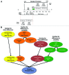
Figure 1. Representation of the workflow of the study. The figure ascribes all the platforms and techniques used in the discovery and the two independent validation sets. The number of samples recruited its time are also depicted in brackets.
None
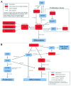
Figure 2. (A) Illustration defining the Greater Promoter region. Using a functional genomic distribution viewpoint we define five CpG genomic locations in relation to their distance to the Transcription Start Site: Proximal promoter, distal promoter, first exon, gene body and intergenic locations. From a CpG content and neighborhood context viewpoint we define four CpG genomic locations in relation to their distance to the nearest CpG Island: CpG Island, CpG Island Shore, CpG Island Shelf, Open Sea and Gene Body. The Greater promoter window is fixed in relation to the TSS. Therefore, the location of CpG Islands will influence the number of significant sequencing reads and 450K probes per gene that are included in our analysis; (B) Workflow for identification of differential methylation in the greater promoter of HNSCC, using next-generation MBD-sequencing and 450K methylation platforms. Schematic description of the analytic pipeline developed to unveil the HNSCC methylome. This pipeline enriches for mean genome-wide differences in CpG methylation as also for genome-wide differences in CpG methylation variability at each chromosomal location, for both, methylation sequencing and methylation 450K array data.
None
Figure 3. Integrative analysis of co-localized promoter methylation and somatic mutations with concurrent expression changes. (A) Methylated genes with fold change differences in expression greater than 2; (B) Genes with co-localized promoter methylation and somatic mutations in HNSCC. Methylation frequency is represented by the red color. Mutation frequency is represented by the blue color. The purple color represents the combined frequency of methylation and mutation events in the Discovery cohort.
None
Figure 4. qMSP results for PAX1, PAX5, ZIC4 and PLCB1. (A) Graphical expression of the logistic regression, Pr (HNSCC = 1) = logit−1 (b0 + b1 x methylation) in tissue from 76 participants with data overlain. The predictor methylation is the qMSP value for each case (1) and each control (0). Cutoff methylation values for PAX1, PAX5 ZIC4 and PLCB1 are shown by the vertical dotted line. Probability of HNSCC is shown in red; (B) Scatterplots of quantitative MSP analysis of candidate genes promoters in the Validation screen cohort, which consisted of 76 HNSCC tumor tissue samples and 19 normal tissue samples obtained from uvulopharyngopalatoplasty (UPPP) procedures performed in non-cancer patients. The relative level of methylated DNA for each gene in each sample was determined as a ratio of MSP for the amplified gene to ACTB and then multiplied by 1000 [(average value of duplicates of gene of interest / average value of duplicates of ACTB) x 1000] for, PAX1, PAX5, ZIC4, and PLCB1. Red line denotes cutoff value; (C) Sensitivity, Specificity and AUC results for qMSP analysis; (D) Receiver Operator Characteristics (ROC) curve for promoter methylation of PAX1, PAX5, ZIC4 and PLCB1 genes in the validation cohort. The figure shows that for this four gene panel the qMSP results have 96% sensitivity, 94% specificity, a 0.97 AUC and a Positive Predictive Value of 98.5%.
None
Figure 5. Proposed genomic and epigenomic interactions in HNSCC: (A) Proposed partial pathway interplay of p53 and PAX5 in HNSCC. Downregulation of PAX5 leads to differentiation. When methylated, PAX5 an upstream target of p53, fails to activate the later which is also silenced due to mutations, and thus DNA repair, Apoptosis, and Growth Arrest pathways are inactive; (B) PAX1-NOTCH1 interplay through crosstalk of Hedgehog and Notch pathways in cell differentiation and proliferation signals. Notch1 induces p21 expression, either directly through the canonical pathway or indirectly through Hes1 and NFAT activation, leading in both cases to cell cycle arrest. Active Notch1 targets either the Hox family or Hes1. Hes1 is active and will block differentiation. The HOX family of transcription factors, downstream targets of Notch signaling, is frequently silenced, thus blocking the activation of PAX1 which is also downregulated in HNSCC and will not promote differentiation. PAX1 expression can also be induced by Shh through Gli2, which is active. Finally, proliferation is promoted through Gli2 interaction with CCND1 and CCND2.

References

    1. Agrawal N, Frederick MJ, Pickering CR, Bettegowda C, Chang K, Li RJ, Fakhry C, Xie TX, Zhang J, Wang J, et al. Exome sequencing of head and neck squamous cell carcinoma reveals inactivating mutations in NOTCH1. Science. 2011;333:1154–7. doi: 10.1126/science.1206923. - DOI - PMC - PubMed
    1. Stransky N, Egloff AM, Tward AD, Kostic AD, Cibulskis K, Sivachenko A, Kryukov GV, Lawrence MS, Sougnez C, McKenna A, et al. The mutational landscape of head and neck squamous cell carcinoma. Science. 2011;333:1157–60. doi: 10.1126/science.1208130. - DOI - PMC - PubMed
    1. Westra WH. The changing face of head and neck cancer in the 21st century: the impact of HPV on the epidemiology and pathology of oral cancer. Head Neck Pathol. 2009;3:78–81. doi: 10.1007/s12105-009-0100-y. - DOI - PMC - PubMed
    1. Kim GB, Wang Z, Liu YY, Stavrou S, Mathias A, Goodwin KJ, Thomas JM, Neville DM. A fold-back single-chain diabody format enhances the bioactivity of an anti-monkey CD3 recombinant diphtheria toxin-based immunotoxin. Protein Eng Des Sel. 2007;20:425–32. doi: 10.1093/protein/gzm040. - DOI - PubMed
    1. Poeta ML, Manola J, Goldwasser MA, Forastiere A, Benoit N, Califano JA, Ridge JA, Goodwin J, Kenady D, Saunders J, et al. TP53 mutations and survival in squamous-cell carcinoma of the head and neck. N Engl J Med. 2007;357:2552–61. doi: 10.1056/NEJMoa073770. - DOI - PMC - PubMed

Publication types

MeSH terms