. 2014 May 27;110(11):2677-87.
doi: 10.1038/bjc.2014.221. Epub 2014 May 1.

Rac1 as a potential therapeutic target for chemo-radioresistant head and neck squamous cell carcinomas (HNSCC)

Affiliations

Rac1 as a potential therapeutic target for chemo-radioresistant head and neck squamous cell carcinomas (HNSCC)

S Skvortsov et al. Br J Cancer. .

Abstract

Background: In order to improve therapy for HNSCC patients, novel methods to predict and combat local and/or distant tumour relapses are urgently needed. This study has been dedicated to the hypothesis that Rac1, a Rho GTPase, is implicated in HNSCC insensitivity to chemo-radiotherapy resulting in tumour recurrence development.

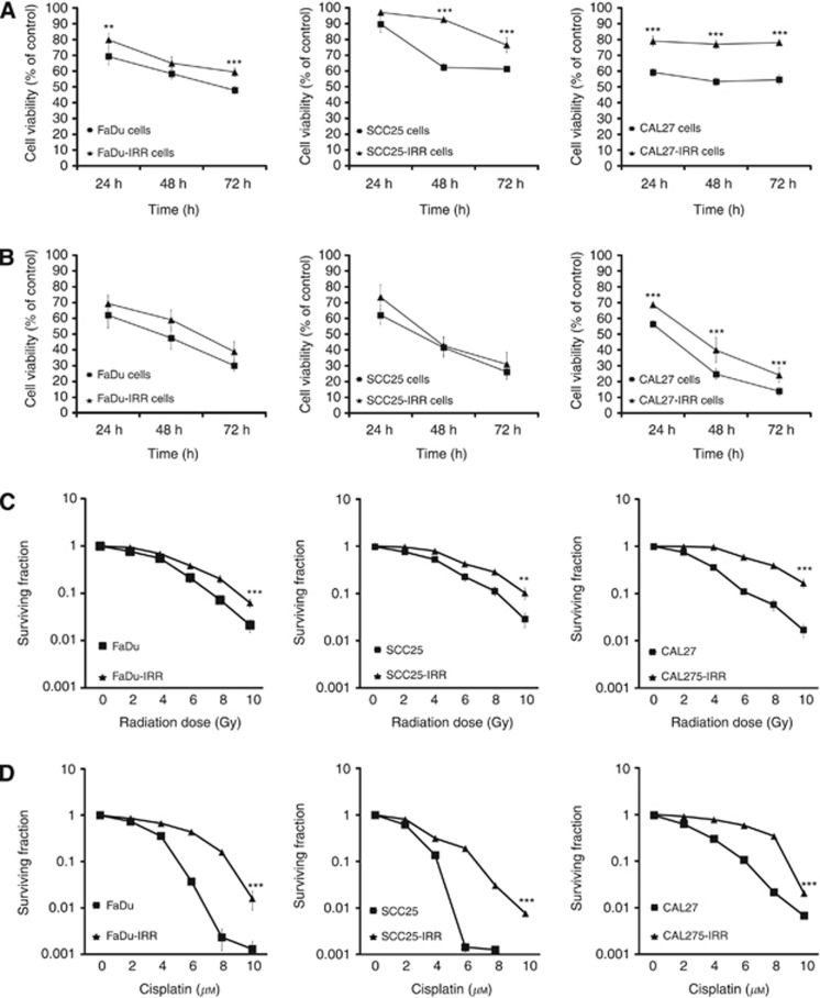
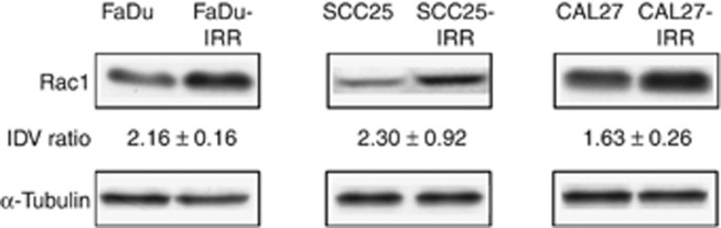
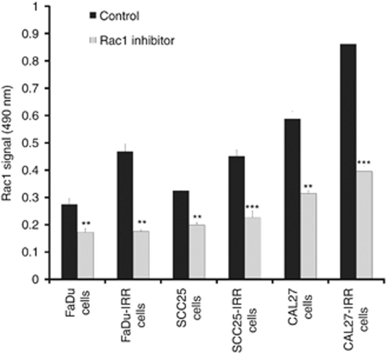
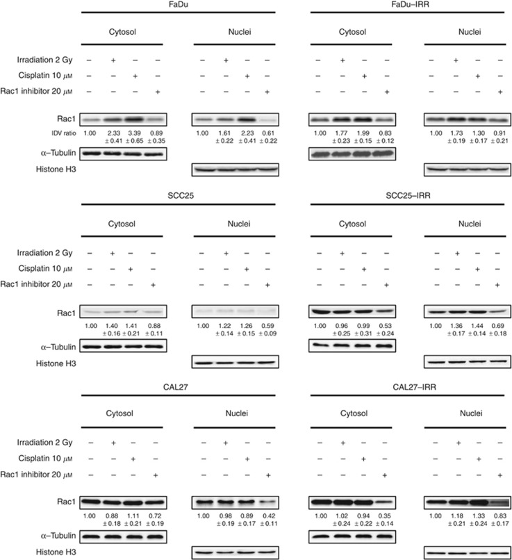
Methods: Parental and radiation-resistant (IRR) HNSCC cells were used to support this hypothesis. All cells were investigated for their sensitivity to ionising radiation and cisplatin, Rac1 activity, its intracellular expression and subcellular localisation. Additionally, tumour tissues obtained from 60 HNSCC patients showing different therapy response were evaluated for intratumoral Rac1 expression.

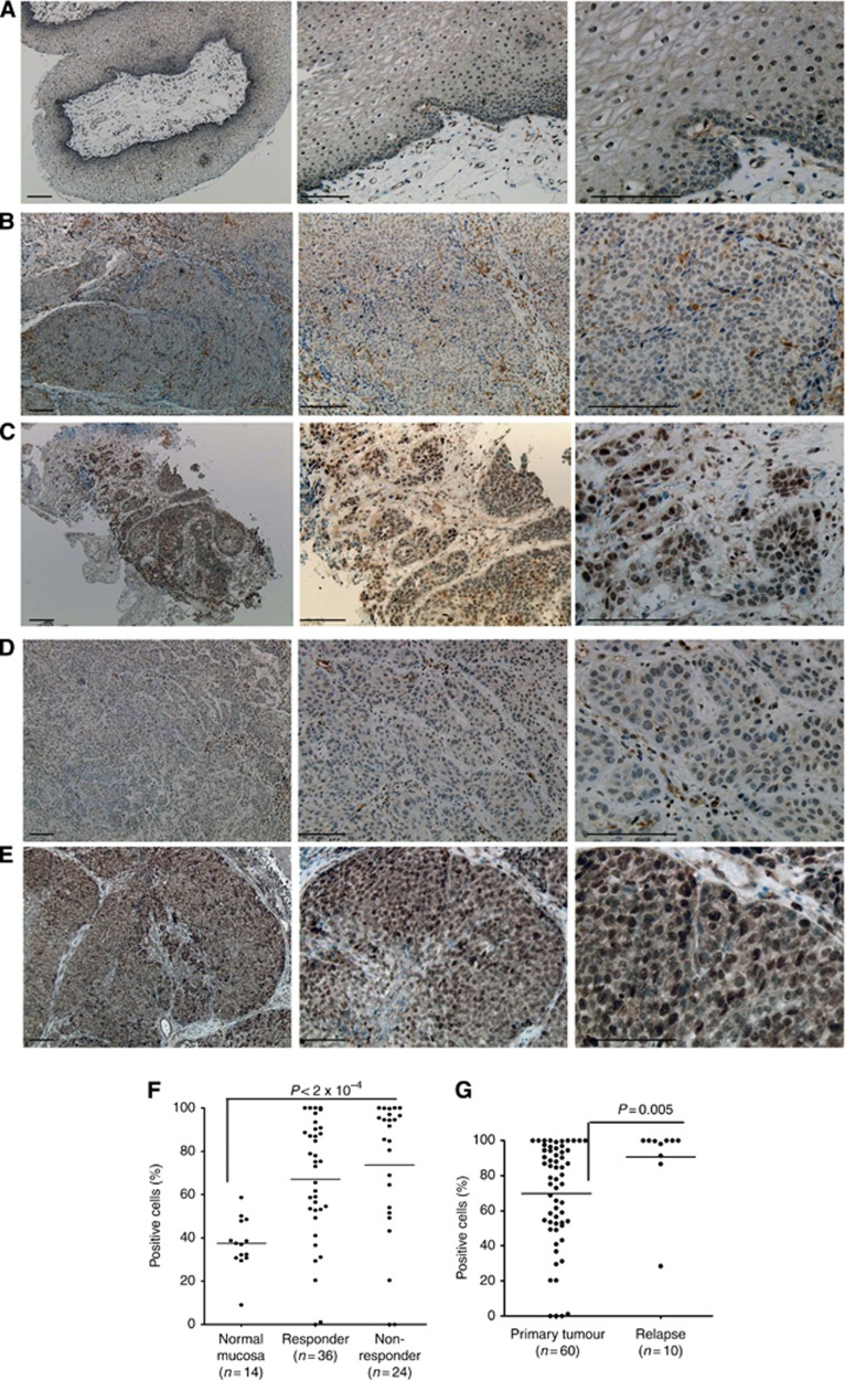
Results: Radiation-resistant IRR cells also revealed resistance to cisplatin accompanied by increased expression, activity and trend towards nuclear translocation of Rac1 protein. Chemical inhibition of Rac1 expression and activity resulted in significant improvement of HNSCC sensitivity to ionising radiation and cisplatin. Preclinical results were confirmed in clinical samples. Although Rac1 was poorly presented in normal mucosa, tumour tissues revealed increased Rac1 expression. The most pronounced Rac1 presence was observed in HNSCC patients with poor early or late responses to chemo-radiotherapy. Tissues taken at recurrence were characterised not only by enhanced Rac1 expression but also increased nuclear Rac1 content.

Conclusions: Increased expression, activity and subcellular localisation of Rac1 could be associated with lower early response rate and higher risk of tumour recurrences in HNSCC patients and warrants further validation in larger independent studies. Inhibition of Rac1 activity can be useful in overcoming treatment resistance and could be proposed for HNSCC patients with primary or secondary chemo-radioresistance.


Figures

Figure 1
Figure 1
Cell viability and clonogenic survival of HNSCC cells in response to radiation and cisplatin exposure. Parental FaDu, SCC25 and CAL27 and appropriate radiation-resistant IRR cells were seeded in six-well plates, treated with ionising radiation at a single dose of 2 Gy (A) or cisplatin at a clinically relevant single dose of 10 μM (B) and then incubated for 72 h. Cell viability and number of cells was evaluated using the Beckman Coulter Vi-CELL AS cell viability analyser. Data are given as mean and s.d. obtained from at least three independent experiments. (C and D) Clonogenic cell survival assay. Colonies were counted on the fourteenth day following radiation (C) or cell exposure to cisplatin (D), and surviving fractions were plotted as a function of dose on a log-linear scale. Error bars indicate s.d. from mean of duplicate measurements from at least four independent experiments. **P<0.01; ***P<0.001.
Figure 2
Figure 2
Rac1 expression in HNSCC cells. Western blotting analysis was done using protein extracts from parental FaDu, SCC25, CAL27 and treatment-resistant FaDu-IRR, SCC25-IRR and CAL27-IRR cells, as described in Materials and methods section. α-Tubulin was used as a loading control. IDV was calculated for each protein band and normalised to the α-tubulin band density after background correction. IDV ratio means fold increase of Rac1 band density in IRR cells to those in parental HNSCC cells. Western blots shown are representative of three independent experiments.
Figure 3
Figure 3
Rac1 activity in parental and treatment-resistant HNSCC cells. Parental and IRR HNSCC cells were serum starved for 24 h and then treated with Rac1 inhibitor (20 μM). Cell lysates were subjected to the G-LISA assay in triplicates, and absorbance of each well was read at 490 nm. Results are shown as mean and s.d. obtained from three independent experiments. **P<0.01; ***P<0.001.
Figure 4
Figure 4
Effects of ionising radiation, cisplatin or Rac1 inhibitor on cytoplasmic and nuclear Rac1 expression in HNSCC cells. Western blotting analysis of Rac1 expression in cytoplasmic and nuclear fractions obtained from parental and treatment-resistant IRR HNSCC cells before and at 24 h after treatment with ionising radiation (2 Gy), cisplatin (10 μM) or Rac1 inhibitor (20 μM). IDV ratio was calculated as ratio between IDV values of treated cells to those in untreated HNSCC. For IDV evaluation, the background corrections have been done for all protein band densities using α-tubulin for cytoplasmic and histone H3 for nuclear fractions.
Figure 5
Figure 5
Rac1 immunohistochemical (IHC) reaction in normal mucosa and in HNSCC tissue samples. IHC representation of Rac1 expression in normal epithelium (A); in radio-chemotherapy HNSCC early responder (B); in chemo-radiotherapy HNSCC non-responder (C), in primary tumour of therapy responder without signs of tumour relapse (D) and in the tumour tissue obtained from relapsed HNSCC patient (E). Left panels of images: × 40 magnification, middle panels: × 100 magnification, right panels: × 200 magnification; bars: 100 μm. (F) Graphical representation of percentage of Rac1-positive cells in normal tissue and tumours obtained from therapy responders and non-responders. (G) Differences in Rac1 expression in tumours from HNSCC patients with positive therapy response and from relapsed HNSCC patients. *P<0.05, **P<0.01, ***P<0.001.

References

    1. Ang KK, Zhang QE, Rosenthal DI, Nguyen-Tan P, Sherman EJ, Weber RS, Galvin JM, Schwartz DL, El-Naggar AK, Gillison ML, Jordan R, List MA, Konski AA, Thorstad WL, Trotti A, Beitler JJ, Garden AS, Spanos WJ, Yom SS, Axelrod RS. A randomized phase III trial (RTOG 0522) of concurrent accelerated radiation plus cisplatin with or without cetuximab for stage III-IV head and neck squamous cell carcinomas (HNC) J Clin Oncol. 2011;29 (suppl:abstr 5500. - PMC - PubMed
    1. Argiris A, Li Y, Forastiere A. Prognostic factors and long-term survivorship in patients with recurrent or metastatic carcinoma of the head and neck. Cancer. 2004;101:2222–2229. - PubMed
    1. Baghi M, Hambek M, Wagenblast J, May A, Gstoettner W, Knecht R. A phase II trial of docetaxel, cisplatin and 5-fluorouracil in patients with recurrent squamous cell carcinoma of the head and neck (SCCHN) Anticancer Res. 2006;26:585–590. - PubMed
    1. Begg AC. Predicting recurrence after radiotherapy in head and neck cancer. Semin Radiat Oncol. 2012;22:108–118. - PubMed
    1. Chen YW, Chu HC, Lin ZS, Shiah WJ, Chou CP, Klimstra DS, Lewis BC. p16 stimulates CDC42-dependent migration of hepatocellular carcinoma cells. PLoS One. 2013;8:e69389. - PMC - PubMed

Publication types

MeSH terms