. 2014 Aug;21(8):1262-74.
doi: 10.1038/cdd.2014.46. Epub 2014 May 2.

ANG II promotes IGF-IIR expression and cardiomyocyte apoptosis by inhibiting HSF1 via JNK activation and SIRT1 degradation

Affiliations

ANG II promotes IGF-IIR expression and cardiomyocyte apoptosis by inhibiting HSF1 via JNK activation and SIRT1 degradation

C-Y Huang et al. Cell Death Differ. 2014 Aug.

Abstract

Hypertension-induced cardiac hypertrophy and apoptosis are major characteristics of early-stage heart failure. Our previous studies found that the activation of insulin-like growth factor receptor II (IGF-IIR) signaling was critical for hypertensive angiotensin II (ANG II)-induced cardiomyocyte apoptosis. However, the detailed mechanism by which ANG II regulates IGF-IIR in heart cells remains elusive. In this study, we found that ANG II activated its downstream kinase JNK to increase IGF-IIR expression through the ANG II receptor angiotensin type 1 receptor. JNK activation subsequently led to sirtuin 1 (SIRT1) degradation via the proteasome, thus preventing SIRT1 from deacetylating heat-shock transcription factor 1 (HSF1). The resulting increase in the acetylation of HSF1 impaired its ability to bind to the IGF-IIR promoter region (nt -748 to -585). HSF1 protected cardiomyocytes by acting as a repressor of IGF-IIR gene expression, and ANG II diminished this HSF1-mediated repression through enhanced acetylation, thus activating the IGF-IIR apoptosis pathway. Taken together, these results suggest that HSF1 represses IGF-IIR gene expression to protect cardiomyocytes. ANG II activates JNK to degrade SIRT1, resulting in HSF1 acetylation, which induces IGF-IIR expression and eventually results in cardiac hypertrophy and apoptosis. HSF1 could be a valuable target for developing treatments for cardiac diseases in hypertensive patients.


Figures

Figure 1
Figure 1
ANG II stimulated IGF-IIR expression to induce apoptosis through the AT1R. (a) H9c2 cells were silenced with the AT1R and AT2R small interfering RNAs (siRNAs) (10 nM) for 24 h. Then, the cells were treated with ANG II (100 nM) for 24 h. The expression of the IGF-IIR mRNA was measured using RT–PCR analysis. (b) H9c2 cells were treated with the AT1R blocker losartan (1 μM) and the AT2R blocker PD123319 (1 μM) with ANG II (100 nM) for 24 h. The expression of the IGF-IIR mRNA was examined by RT–PCR analysis. (c) H9c2 cells were transfected with pGL4-IGF-IIR (nt −1249∼+11) together with an siRNA or overexpression plasmid for 24 h. The cells were then treated with ANG II (100 nM) and assayed for luciferase activity. The results are shown as the means±S.D. of three independent experiments. *P<0.05, represents a significant increase compared with the untreated control. ###P<0.001, represents a significant decrease compared with the untreated control. (d) H9c2 cells were treated with losartan or PD123319 with ANG II for 24 h and then assayed for luciferase activity. The results are shown as the means±S.D. of three independent experiments. **P<0.01, represents a significant increase compared with the untreated control. ***P<0.001, represents a significant increase compared with the untreated control. ###P<0.001, represents a significant decrease compared with the untreated control. (e) H9c2 cells were transfected with the AT1R siRNA for 24 h and then treated with ANG II (100 nM) for 24 h or co-treated with the AT1R blocker losartan and ANG II for 24 h. IGF-IIR and AT1R levels were measured by immunoblotting. (f) H9c2 cells were transfected with Flag-AT1R for 24 h and then treated with ANG II (100 nM). The levels of IGF-IIR, caspase-3 and AT1R were measured by immunoblotting. (g) H9c2 cells were transfected with siAT1R, siAT2R and Flag-AT1R or treated with Losartan (AT1R blocker) and PD123319 (AT2R blocker) for 24 h, and then challenged with ANG II for 24 h. The membrane IGF-IIR was detected by ELISA. *P<0.5, represents a significant increase compared with the untreated control. ***P<0.01, represents a significant increase compared with the untreated control. ###P<0.001, represents a significant decrease compared with the untreated control. All the results are representative of three independent experiments
Figure 2
Figure 2
ANG II enhanced IGF-IIR expression via c-Jun N-terminal kinase (JNK), extracellular-signal-regulated kinase (ERK) and phosphatidylinositol 3-kinase (PI3K). (a) H9c2 cells were co-treated with a PI3K inhibitor (LY294002, 10 μM), MEK inhibitor (U0126, 30 μM), ERK inhibitor (PD98059, 10 μM), JNK inhibitor (SP600125, 20 μM) or p38 inhibitor (SB203580, 10 μM) together with ANG II (100 nM). IGF-IIR expression was measured by immunoblotting. (b) H9c2 cells were treated with small interfering RNAs (siRNAs) (10 nM) for PI3K, ERK and JNK individually. After 24 h, the cells were exposed to ANG II (100 nM). The levels of IGF-IIR, PI3K, phosphorylated (p)-ERK and p-JNK1/2 were examined by immunoblotting. (c) The membrane IGF-IIR of H9c2 cells was detected by ELISA following treatment with kinase inhibitors and ANG II. **P<0.01, represents a significant increase compared with the untreated control. ###P<0.001, represents a significant decrease compared with the untreated control. (d) H9c2 cells transfected with pGL4-IGF-IIR (nt −1249 to +11) for 24 h and then treated with a distinct kinase inhibitor and ANG II for 24 h. The cells were assayed for luciferase activity. The results are shown as the means±S.D. of three independent experiments. *P<0.05, represents a significant increase compared with the untreated control. **P<0.01, represents significant increase comparison with untreated control. ***P<0.001, represents a significant increase compared with the untreated control. ##P<0.01, represents a significant decrease compared with the untreated control. ###P<0.001, represents a significant decrease compared with the untreated control. The results are representative of three independent experiments
Figure 3
Figure 3
c-Jun N-terminal kinase (JNK) activation upregulated the expression of IGF-IIR, which translocated to the membrane. (a) H9c2 cells were treated with increasing concentrations of the JNK activator anisomycin (0, 50, 100 and 200 nM) combined with ANG II. The lysates were analyzed by immunoblotting with IGF-IIR, JNK and caspase-3 antibodies. (b and c) After treatment with anisomycin (JNK activator), SP600125 (JNK inhibitor) and ANG II for 24 h, the cells were fixed with 4% paraformaldehyde. Immunofluorescence was measured using anti-IGF-IIR antibodies. White arrows indicate the IGF-IIR protein. *P<0.05 represents a significant increase compared with untreated control. **P<0.01 represents a significant increase in comparison with untreated control. ##P<0.01 represents a significant decrease of membrane IGF-IIR in comparison with ANG II-treated cells. All the blots and micrographs are representative of two to three sets of separate experiments
Figure 4
Figure 4
HSF1 suppressed IGF-IIR expression by binding to its promoter region (nt −748 to −585). (a) Schematic diagram of the IGF-IIR promoter deletion mutations. (b) H9c2 cells were transfected with different IGF-IIR luciferase constructs for 24 h and then treated with ANG II for 24 h. Cell lysates were assayed for luciferase activity. *P<0.05 represents a significant increase in comparison with untreated control. (c) Sequence analysis and transcription factor site prediction identified one putative HSF1 binding element at the IGF-IIR promoter (nt −733 to −706). (d) After treatment with ANG II, the H9c2 cells were lysed and analyzed by ChIP. HSF1 binding to the IGF-IIR promoter was quantified using PCR. (e) HSF1 was silenced with small interfering RNA (siRNA) (10 nM) and transfected with pGL4-IGF-IIR (nt−1249∼+11) for 24 h, followed by exposure to ANG II (100 nM) for 24 h. The lysates were analyzed for luciferase activity. *P<0.05 represents a significant increase in comparison with untreated control. **P<0.01 represents a significant increase in comparison with untreated control. (f) After treatment with the HSF1 inhibitor triptolide (1 μM) and the HSF1 siRNA (10 nM) for 24 h, H9c2 cells were challenged with ANG II (100 nM). IGF-IIR and HSF1 levels were measured by immunoblotting. (g) H9c2 cells were silenced with the HSF1 siRNA (10 nM) or the HSF1 inhibitor triptolide (1 μM) for 24 h and then challenged with ANG II (100 nM) for 24 h. The membrane levels of IGF-IIR were analyzed by ELISA. **P<0.01 represents a significant increase in comparison with untreated control. ***P<0.001 represents a significant increase in comparison with untreated control. ###P<0.001 represents a significant decrease in comparison with untreated control. All the blots are representative of two to three sets of independent experiments
Figure 5
Figure 5
The ability of HSF1 to repress IGF-IIR was reduced by ANG II via posttranslational modification. (a) H9c2 cells were treated with ANG II, and the cell lysates were fractionated into cytosolic and nuclear proteins. HSF1 (both the active and the Ser303-phosphorylated inactive form) were analyzed by immunoblotting. α-Tubulin and histone deacetylase (HDAC) served as the cytosolic and nuclear loading controls. (b and c) H9c2 cells were cultured on slides and treated with ANG II for 24 h. Immunofluorescence analysis was performed with the anti-HSF1 antibody and Alexa Fluor 488. The nuclear compartment was stained with 4′,6-diamidino-2-phenylindole (DAPI), and the cells were visualized under a confocal microscope. *P<0.05, represents a significant increase compared with the untreated control. (d) H9c2 cells were transfected with Flag-HSF1 for 24 h and treated with ANG II for 24 h. Then, 500 μg of cell lysate was immunoprecipitated with anti-Flag antibodies and detected with anti-acetyl-lysine antibodies by immunoblotting. (e) H9c2 cells were treated with ANG II for 24 h. Then, 500 μg of cell lysate was immunoprecipitated with anti-HSF1 and anti-acetyl antibodies and analyzed by immunoblotting. (f) H9c2 cells were treated with ANG II for 24 h. The HSF1 acetylated residue, lysine 80, is conserved in human, mouse and rat. SIRT1 levels were assayed by immunoblotting
Figure 6
Figure 6
JNK induced IGF-IIR expression by degrading SIRT1 via the proteasome to impair HSF1 deacetylation. (a) H9c2 cells were silenced with the SIRT1 small interfering RNA (siRNA) for 24 h and analyzed by RT-PCR and immunoblotting. (b) After transfection with SIRT1-wt or SIRT1-H363Y (deacetylase defective mutant) for 24 h, H9c2 cardiomyoblast cells were treated with ANG II (100 nM) alone or combined with the SIRT1 activator RSV (50 mM) for 24 h. The expression of IGF-IIR was assayed by immunoblotting. (c) H9c2 cells were transfected with SIRT1-wt (wild-type) and treated with ANG II. The lysates were analyzed by immunoprecipitation and immunoblotting. (d) H9c2 cells were transfected with Flag-HSF1 for 24 h and treated with ANG II alone or combined with the SIRT1 inhibitor NAM (10 mM) for 24 h. The expression of IGF-IIR and SIRT1 was examined by immunoblotting. (e) H9c2 cells were treated with ANG II alone or combined with NAM (10 mM) and SP600125 (20 μM) and then analyzed by immunoprecipitation and immunoblotting. The acetylation of HSF1 was detected using an anti-acetyl-lysine antibody. (f) H9c2 cells were treated with ANG II or anisomycin for 15 h, followed by the addition of the proteasome inhibitor MG132 (10 mM) for 9 h. The expression of SIRT1 was assayed by immunoblotting. (g) H9c2 cells were transfected with hemagglutinin (HA)-ubiquitin for 24 h, and then ANG II and MG132 were added. The lysates were immunoprecipitated with specific SIRT1 antibodies and analyzed by immunoblotting
Figure 7
Figure 7
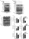
JNK enhanced SIRT1 degradation to activate IGF-IIR- induced hypertrophy and apoptosis in NRVMs and SHR hearts. (a) After treatment with ANG II (100 nM), anisomycin (50 ng/ml), RSV (50 mM), NAM (10 mM), or anisomycin/NAM for 24 h, H9c2 cell lysates were analyzed by immunoblotting. (b) NRVM primary cells were first transfected with Flag-HSF1 or the HSF1 siRNA (10 nM) for 24 h and then treated with ANG II (100 nM) and anisomycin (50 ng/ml), SP600125 (20 μM) or NAM (10 mM) for 24 h. *A non-specific band. (c) Rat heart tissues were homogenized and extracted for analysis. The expression of IGF-IIR, JNK1/2, SIRT1, HSF1, active caspase-3 and BNP were measured by western blot analysis. (d) The relative expression of IGF-IIR, pJNK1, SIRT1, HSF1, Active Casp.3 and BNP was analyzed by Image J and normalized with the WKY group. *P<0.05 represents a significant increase in comparison with untreated control. **P<0.01 represents a significant increase in comparison with untreated control. ***P<0.001 represents a significant increase in comparison with untreated control. #P<0.05 represents a significant decrease in comparison with untreated control
Figure 8
Figure 8
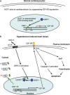
A proposed pathway by which ANG II releases HSF1 from the DNA via the AT1R-JNK1/2-SIRT1 pathway, thus upregulating IGF-IIR expression in cardiomyocytes and hypertensive hearts. (a) Under normal conditions, HSF1 acts as repressor to suppress IGF-IIR by binding to its HSE in the IGF-IIR promoter region (nt −733 to −706). Therefore, HSF1 protects cardiomyocytes by inhibiting IGF-IIR-induced apoptosis. (b) Once ANG II binds to its receptor AT1R, the downstream kinase JNK1/2 is activated to reduce SIRT1 levels via ubiquitin degradation. Without the HSF1 deacetylase SIRT1, the acetylated HSF1 accumulates; at the same time, the levels of the cytosolic inactive HSF1Ser303-phosphorylated protein increase. Therefore, HSF1 cannot bind to the IGF-IIR promoter and suppress its expression. IGF-IIR is upregulated and translocates into the surface membrane to sensitize the serum IGF-II, which activates its downstream protein Gαq to induce hypertrophy and apoptosis. Finally, this phenomenon leads to hypertension-induced heart failure

References

    1. George AJ, Thomas WG, Hannan RD. The renin–angiotensin system and cancer: old dog, new tricks. Nat Rev Cancer. 2010;10:745–759. - PubMed
    1. Herichova I, Szantoova K. Renin–angiotensin system: upgrade of recent knowledge and perspectives. Endocr Regul. 2013;47:39–52. - PubMed
    1. Mehta PK, Griendling KK. Angiotensin II cell signaling: physiological and pathological effects in the cardiovascular system. Am J Physiol Cell Physiol. 2007;292:C82–C97. - PubMed
    1. Hunyady L, Catt KJ. Pleiotropic AT1 receptor signaling pathways mediating physiological and pathogenic actions of angiotensin II. Mol Endocrinol. 2006;20:953–970. - PubMed
    1. Braulke T. Type-2 IGF receptor: a multi-ligand binding protein. Horm Metab Res. 1999;31 (2–3:242–246. - PubMed

Publication types

LinkOut - more resources