. 2014 Sep;21(9):1460-8.
doi: 10.1038/cdd.2014.63. Epub 2014 May 2.

The TORC1 inhibitors Nprl2 and Nprl3 mediate an adaptive response to amino-acid starvation in Drosophila

Affiliations

The TORC1 inhibitors Nprl2 and Nprl3 mediate an adaptive response to amino-acid starvation in Drosophila

Y Wei et al. Cell Death Differ. 2014 Sep.

Abstract

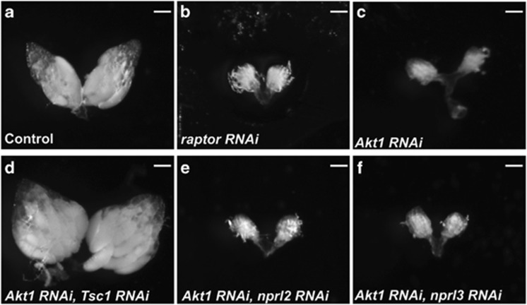
Target of rapamycin complex 1 (TORC1) is a master regulator of metabolism in eukaryotes that integrates information from multiple upstream signaling pathways. In yeast, the Nitrogen permease regulators 2 and 3 (Npr2 and Npr3) mediate an essential response to amino-acid limitation upstream of TORC1. In mammals, the Npr2 ortholog, Nprl2, is a putative tumor suppressor gene that inhibits cell growth and enhances sensitivity to numerous anticancer drugs including cisplatin. However, the precise role of Nprl2 and Nprl3 in the regulation of metabolism in metazoans remains poorly defined. Here we demonstrate that the central importance of Nprl2 and Nprl3 in the response to amino-acid starvation has been conserved from single celled to multicellular animals. We find that in Drosophila Nprl2 and Nprl3 physically interact and are targeted to lysosomes and autolysosomes. Using oogenesis as a model system, we show that Nprl2 and Nprl3 inhibit TORC1 signaling in the female germline in response to amino-acid starvation. Moreover, the inhibition TORC1 by Nprl2/3 is critical to the preservation of female fertility during times of protein scarcity. In young egg chambers the failure to downregulate TORC1 in response to amino-acid limitation triggers apoptosis. Thus, our data suggest the presence of a metabolic checkpoint that initiates a cell death program when TORC1 activity remains inappropriately high during periods of amino-acid and/or nutrient scarcity in oogenesis. Finally, we demonstrate that Nprl2/3 work in concert with the TORC1 inhibitors Tsc1/2 to fine tune TORC1 activity during oogenesis and that Tsc1 is a critical downstream effector of Akt1 in the female germline.


Figures

Figure 1
Figure 1
Nprl2 and Nprl3 inhibit TORC1 activity in Drosophila. (a) S2 cells were co-transfected with HA-tagged Nprl2 and V5-tagged Nprl3 or lacZ (control) plasmids. Cells were lysed and immunoprecipitated using an anti-V5 antibody. Cell lysates (inputs) and immunoprecipitates (IP) were detected by western blot using anti-HA and anti-V5 antibodies. (b) S2 cells were treated with GFP, nprl2 or nprl3 dsRNA for 4 days and then cultured in Schneider's medium plus 10% FBS (AA+) or amino-acid-free Schneider's medium (AA-) for 60 min. The protein levels of p-4E-BP, total 4E-BP and α-Tubulin were determined by western blot. Similar western Blot results were observed in three independent experiments. (c) S2 cells were treated with GFP, nprl2 or nprl3 dsRNA for 4 days and then cultured in amino-acid-free Schneider's medium plus 10% FBS for 24 h. Cell size in G1 phase was normalized to GFP dsRNA control. Error bars represent S.D. values from three independent experiments. *P<0.05; **P<0.01
Figure 2
Figure 2
Nprl2 and Nprl3 localize to lysosomes, autolysosomes. (ao and a'o') Live cell imaging of Drosophila egg chambers from females cultured on fly media with wet yeast (fed, ao) or 20% sucrose (starved, a'o'). (ac and a'c') GFP-Nprl2 co-localizes with LysoTracker Red. (df and d'f') Nprl3-mCherry co-localizes with LysoTracker Green. (gi and g'i') GFP-Nprl2 co-localizes with mCherry-Atg8. (jl and j'l') Nprl3-mCherry co-localizes with GFP-LAMP1. (mo and m'o') GFP-Nprl2 co-localizes with Nprl3-mCherry. Note that Nprl2 and Nprl3 localize to autolysosomes (arrow; AL, autolysosome). In addition, Nprl2 and Nprl3 are found in nuclei (arrowhead; N, nuclei). The MTD-GAL4 driver was used to drive expression of GFP-Nprl2, mCherry-Atg8 and Nprl3-mCherry. GFP-LAMP1 was expressed with a tubulin promoter. Bar, 10 μm
Figure 3
Figure 3
Nprl2 and Nprl3 protect young egg chambers during amino-acid starvation. (ai) Females were cultured on 20% sucrose (AA-) for 5 days. (ac) MTD>mCherry RNAi, (df) MTD>nprl2 RNAi-1 and (gi) MTD>nprl3 RNAi-1 egg chambers, labeled with DAPI (blue) and cleaved Caspase-3 (red). Note that the egg chambers with condensed DNA staining (pyknotic nuclei) were also cleaved Caspase-3 positive (arrows). Bar, 10 μm. (j) Females were cultured on 20% sucrose (AA-) for indicated number of days. Percentages of dying egg chambers (stage 2–7) were recorded for each genotype. Error bars represent S.D. of three independent experiments. The numbers above bars represent the number of young egg chambers (stages 2–7) examined. (k) Females were cultured on 20% sucrose for 7 days and then transferred to fly media containing wet yeast. Data show eggs laid after the cessation of starvation. Error bars represent S.D. of three independent experiments, where each experiment examined greater than 200 ovaries. *P<0.05; **P<0.01
Figure 4
Figure 4
TORC1 inhibition promotes young egg chambers survival during amino-acid starvation. (a) MTD>mcherry RNAi females were cultured on 20% sucrose plus or minus rapamycin for 9 days. (b) MTD>nprl2 RNAi females were cultured on 20% sucrose plus or minus rapamycin for 9 days. (c) MTD>nprl3 RNAi females were cultured on 20% sucrose plus or minus rapamycin for 5 days. Data represent the percentages of dead or dying egg chambers (stage 2-7). (d) Females were cultured on 20% sucrose plus rapamycin for 7 days and then transferred to fly media containing wet yeast. Data show eggs laid numbers after the cessation of starvation. (e, f) MTD>mCherry RNAi and MTD>Tsc1 RNAi females were cultered on 20% sucrose (AA-) for 5 days. The young egg chambers were labeled with DAPI (blue) and cleaved Caspase-3 (red). Bar, 10 μm. (f) MTD>Tsc1 RNAi females were cultured on 20% sucrose (AA-) for indicated days. Percentages of dead egg chambers (stage 2–7) were recorded. Error bars represent S.D. *P<0.05; **P<0.01. In ac and g, the numbers above bars represent the number of young egg chambers (stages 2–7) examined
Figure 5
Figure 5
The TORC1 inhibitor Tsc1 is downstream of Akt1 in the female germline. (a) nanos-GAL4; UAS-mCherry RNAi, (b) nanos-GAL4; UAS-raptor RNAi, (c) nanos-GAL4; UAS-Akt1 RNAi, (d) nanos-GAL4; UAS-Akt1 RNAi/UAS-Tsc1 RNAi, (e) nanos-GAL4; UAS-Akt1 RNAi/UAS-nprl2 RNAi and (f) nanos-GAL4; UAS-Akt1 RNAi/UAS-nprl3 RNAi flies were cultured on standard fly media with wet yeast 2 days before dissection. The germline-specific driver nanos-GAL4 was used to drive expression of the RNAi constructs. (a, d) Hundred percent of the ovaries examined contained mature (stage 13–14) egg chambers. (b, c, e, f) Zero percent of the ovaries examined contained egg chambers beyond stage 5: (af) N>50 ovaries. Bar, 50 μm
Figure 6
Figure 6
The TORC1 inhibitors Nprl2/3 and Tsc1/2 prevent apoptosis during amino-acid starvation in young egg chambers. (a) In the presence of amino acids, the TORC1 inhibitors Nprl2/3 are inactive while the insulin pathway inhibits the activity of Tsc1/2. Thus, TORC1 activity is high driving anabolic metabolism and growth. (b) In the absence of amino acids, Nprl2/3 functions to inhibit TORC1 activity. Amino-acid starvation also results in reduced insulin signaling, leading to activation of the Tsc1/2 complex due to the reduced activity of its inhibitor Akt1. Together Nprl2/3 and Tsc1/2 sufficiently inhibit TORC1 activity to induce an essential stress response (c) In nprl2 or nprl3 knockdowns, cells fail to adequately downregulate TORC1 activity in response to amino-acid starvation triggering apoptosis. (d) In Tsc1 knockdowns, TORC1 activity also remains inappropriately high during amino-acid starvation triggering apoptosis

References

    1. King RC. The meiotic behavior of the Drosophila oocyte. Int Rev Cytol. 1970;28:125–168. - PubMed
    1. Drummond-Barbosa D, Spradling AC. Stem cells and their progeny respond to nutritional changes during Drosophila oogenesis. Dev Biol. 2001;231:265–278. - PubMed
    1. LaFever L, Drummond-Barbosa D. Direct control of germline stem cell division and cyst growth by neural insulin in Drosophila. Science. 2005;309:1071–1073. - PubMed
    1. Spradling AC.In: Bate M, Martinez-Arias A (eds). The Development of Drosophila melanogaster Cold Spring Harbor Laboratory Press: NY, USA; 19931–70.
    1. Shimada Y, Burn KM, Niwa R, Cooley L. Reversible response of protein localization and microtubule organization to nutrient stress during Drosophila early oogenesis. Dev Biol. 2011;355:250–262. - PMC - PubMed

Publication types

MeSH terms