. 2014 May 1;5(5):e1201.
doi: 10.1038/cddis.2014.150.

Reversion of apoptotic resistance of TP53-mutated Burkitt lymphoma B-cells to spindle poisons by exogenous activation of JNK and p38 MAP kinases

Affiliations

Reversion of apoptotic resistance of TP53-mutated Burkitt lymphoma B-cells to spindle poisons by exogenous activation of JNK and p38 MAP kinases

M Farhat et al. Cell Death Dis. .

Abstract

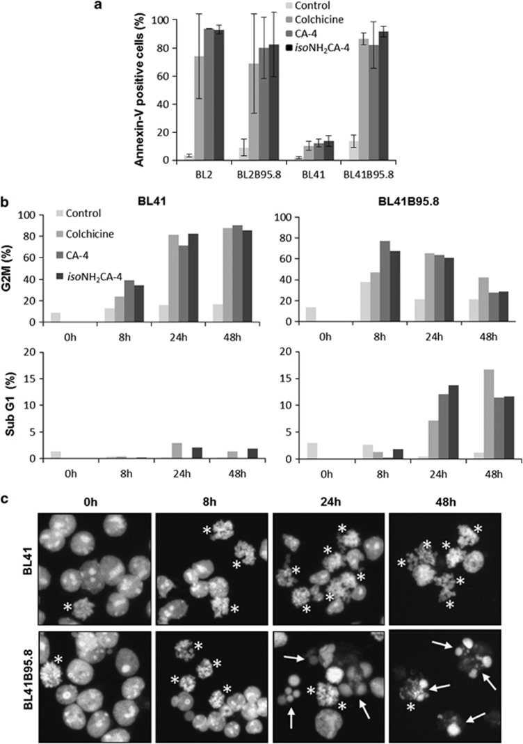
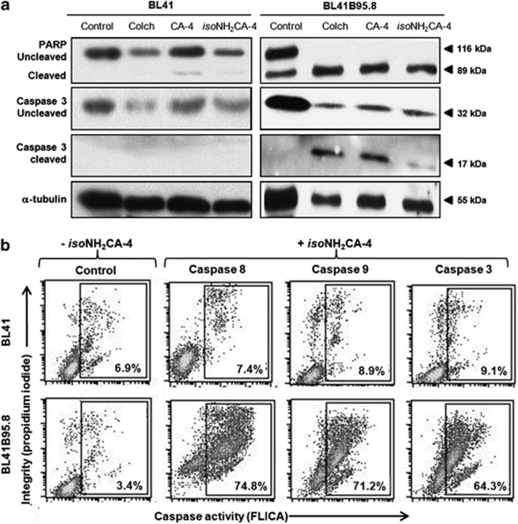
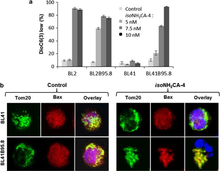
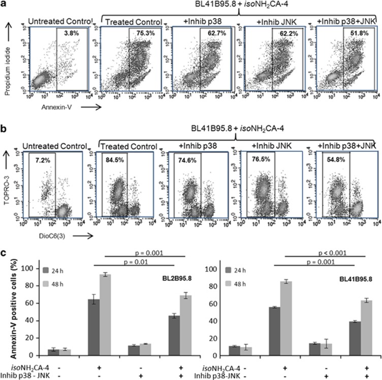
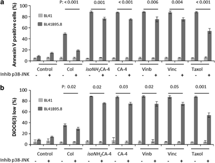
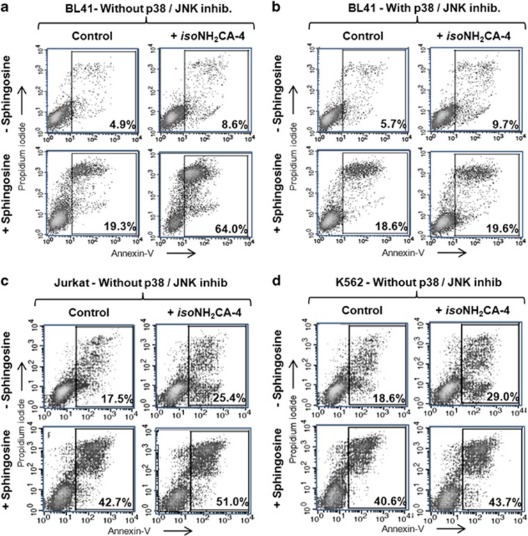
Defects in apoptosis are frequently the cause of cancer emergence, as well as cellular resistance to chemotherapy. These phenotypes may be due to mutations of the tumor suppressor TP53 gene. In this study, we examined the effect of various mitotic spindle poisons, including the new isocombretastatin derivative isoNH2CA-4 (a tubulin-destabilizing molecule, considered to bind to the colchicine site by analogy with combretastatin A-4), on BL (Burkitt lymphoma) cells. We found that resistance to spindle poison-induced apoptosis could be reverted in tumor protein p53 (TP53)-mutated cells by EBV (Epstein Barr virus) infection. This reversion was due to restoration of the intrinsic apoptotic pathway, as assessed by relocation of the pro-apoptotic molecule Bax to mitochondria, loss of mitochondrial integrity and activation of the caspase cascade with PARP (poly ADP ribose polymerase) cleavage. EBV sensitized TP53-mutated BL cells to all spindle poisons tested, including vincristine and taxol, an effect that was systematically downmodulated by pretreatment of cells with inhibitors of p38 and c-Jun N-terminal kinase (JNK) mitogen-activated protein kinases. Exogenous activation of p38 and JNK pathways by dihydrosphingosine reverted resistance of TP53-mutated BL cells to spindle poisons. Dihydrosphingosine treatment of TP53-deficient Jurkat and K562 cell lines was also able to induce cell death. We conclude that activation of p38 and JNK pathways may revert resistance of TP53-mutated cells to spindle poisons. This opens new perspectives for developing alternative therapeutic strategies when the TP53 gene is inactivated.


Figures

Figure 1
Figure 1
EBV renders TP53-mutated cells permissive to spindle poison-induced apoptosis. (a) Cells were treated with colchicine (20 nM), CA-4 (10 nM) or isoNH2CA-4 (5 nM) for 72 h (a and b). (a) Apoptosis induction was evaluated by flow cytometry after labeling of external phosphatidyl serine residues (Annexin-V-positive cells) on BL2- and TP53-mutated BL41 cell lines as well as their EBV-infected counterparts (BL2B95.8 and BL41B95.8). (b) Percentage of cells in G2M (M, mitosis) and sub-G1 phases was evaluated by flow cytometry cell cycle analysis (propidium iodide staining of total DNA content). (c) The same isoNH2CA-4-treated cells were also analyzed by epifluorescence microscopy. Cells in M are indicated by *. Cells with fragmented nuclei are indicated by →. Results are representative of three independent experiments
Figure 2
Figure 2
Apoptosis induced by spindle poisons in EBV-infected BL41B95.8 cells is caspase 3, 8 and 9 dependent. BL41 and BL41B95.8 cells were treated with colchicine (20 nM), CA-4 (10 nM) or isoNH2CA-4 (5 nM) for 24 h. (a) PARP and caspase 3 cleavage as well as α-tubulin (loading control) were analyzed by western blot. (b) Detection of caspase 8, 9 and 3 activation was evaluated by flow cytometry using the caspase-specific FLICA assay. Results are representative of three independent experiments
Figure 3
Figure 3
Mitochondria have a pivotal role in apoptosis induced by isoNH2CA-4 in EBV-infected BL41B95.8 cells. Cells were treated with 5, 7.5 or 10 nM isoNH2CA-4 for 24 h. (a) Mitochondrial integrity was evaluated by flow cytometry using DiOC6(3) staining of BL2- and TP53-mutated BL41 cell lines and their EBV-infected counterparts (BL2B95.8 and BL41B95.8). (b) For BL41 and BL41B95.8 cells treated with 10 nM isoNH2CA-4 for 24 h, colocalization of Bax with the mitochondrial marker Tom20 was studied by confocal microscopy (original magnification × 630). Results are the mean of three independent experiments
Figure 4
Figure 4
Apoptosis is induced in EBV-infected BL2 cells treated with isoNH2CA-4 after TP53 downregulation. Downregulation of TP53 expression was obtained (+siRNAp53) or not (−siRNAp53) by transfection of BL2 (a) and BL2B95.8 (b) cells with either small interfering RNA (siRNA) against TP53 mRNA or scramble siRNA, respectively. Apoptosis induction as evaluated by flow cytometry (Annexin-V labeling). Cells were treated with 10 nM of IsoNH2CA-4. Results are representative of three independent experiments
Figure 5
Figure 5
p38 and JNK MAP kinase inhibition decreases apoptosis induced by isoNH2CA-4 in EBV-infected BL41B95.8 cells. Cells were treated with 10 nM isoNH2CA-4 with or without 1 h pretreatment with the p38 inhibitor SB203580 (25 μM), the JNK inhibitor SP600125 (7.5 nM) or both. (a and b) An example of apoptosis induction and mitochondrial loss of integrity in BL41B95.8 cells as assessed on flow cytometry biparametric histogram after annexin-V and propidium iodide (a) or DiOC6(3) and propidium iodide (b) labeling. Percentages of apoptotic cells are indicated in each graph. (c) Percentages of BL2B95.8 (left panel) and BL41.B95.8 (right panel) annexin-V-positive cells in presence (+) or absence (−) of isoNH2CA-4 and/or both p38 and JNK inhibitors at 24 and 48 h. Results are representative of three independent experiments
Figure 6
Figure 6
p38 and JNK inhibitors counteract apoptosis induced by any class of spindle poisons in EBV-positive BL41B95.8 cells. BL41 and BL41B95.8 cells were treated with actin-stabilizing (Taxol) or with actin-destabilizing drugs specific for the colchicine site (colchicine: 30 nM Col; 10 nM (isoNH2CA-4, isoCA-4); 20 nM CA-4) or the vinca site (vinblastine: 25 nM Vinb; vincristine: 25 nM Vinc) for 24 h. Cells were additionally pretreated (+) or not (−) for 1 h with the p38 inhibitor SB203580 (25 μM) and the JNK inhibitor SP600125 (7.5 nM). (a) Percentages of BL41 and BL41B95.8 annexin-V-positive cells assessed by flow cytometry. (b) Percentages of DiOC6(3) negative cells, assessed by flow cytometry. These results correspond to at least three independent experiments
Figure 7
Figure 7
Dihydrosphingosine renders TP53-mutated and EBV-negative BL41 cells permissive to apoptosis. BL41 (a and b), Jurkat (c) and K562 (d) cells were pretreated or not for 1 h with dihydrosphingosine (7.5 μM) and then treated or not with 10 nM isoNH2CA-4 for 24 h. Apoptosis induction was evaluated by flow cytometry, after annexin-V and propidium iodide staining. Cells were pretreated with dihydrosphingosine in absence (a, c and d) or in presence (b) of both p38 and JNK inhibitors. Percentages of apoptotic cells are indicated in each graph. Results are representative of three independent experiments

Similar articles

Cited by

References

    1. Henson PM, Bratton DL, Fadok VA. Apoptotic cell removal. Curr Biol. 2001;11:R795–R805. - PubMed
    1. Green DR, Kroemer G. The pathophysiology of mitochondrial cell death. Science. 2004;305:626–629. - PubMed
    1. Kroemer G, Galluzzi L, Brenner C. Mitochondrial membrane permeabilization in cell death. Physiol Rev. 2007;87:99–163. - PubMed
    1. LaCasse EC, Mahoney DJ, Cheung HH, Plenchette S, Baird S, Korneluk RG. IAP-targeted therapies for cancer. Oncogene. 2008;27:6252–6275. - PubMed
    1. Matson DR, Stukenberg PT. Spindle poisons and cell fate: a tale of two pathways. Mol Interv. 2011;11:141–150. - PMC - PubMed

MeSH terms

Substances