Review

An official American Thoracic Society/European Respiratory Society statement: update on limb muscle dysfunction in chronic obstructive pulmonary disease

François Maltais et al. Am J Respir Crit Care Med. .

Abstract

Background: Limb muscle dysfunction is prevalent in chronic obstructive pulmonary disease (COPD) and it has important clinical implications, such as reduced exercise tolerance, quality of life, and even survival. Since the previous American Thoracic Society/European Respiratory Society (ATS/ERS) statement on limb muscle dysfunction, important progress has been made on the characterization of this problem and on our understanding of its pathophysiology and clinical implications.

Purpose: The purpose of this document is to update the 1999 ATS/ERS statement on limb muscle dysfunction in COPD.

Methods: An interdisciplinary committee of experts from the ATS and ERS Pulmonary Rehabilitation and Clinical Problems assemblies determined that the scope of this document should be limited to limb muscles. Committee members conducted focused reviews of the literature on several topics. A librarian also performed a literature search. An ATS methodologist provided advice to the committee, ensuring that the methodological approach was consistent with ATS standards.

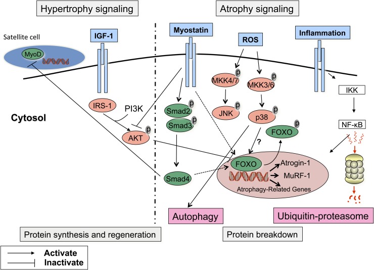
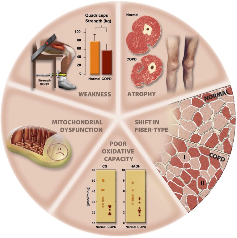
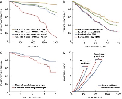
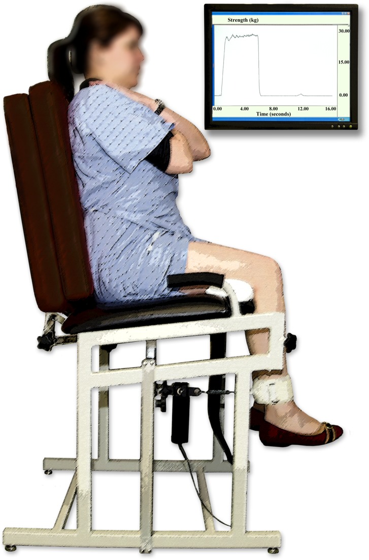
Results: We identified important advances in our understanding of the extent and nature of the structural alterations in limb muscles in patients with COPD. Since the last update, landmark studies were published on the mechanisms of development of limb muscle dysfunction in COPD and on the treatment of this condition. We now have a better understanding of the clinical implications of limb muscle dysfunction. Although exercise training is the most potent intervention to address this condition, other therapies, such as neuromuscular electrical stimulation, are emerging. Assessment of limb muscle function can identify patients who are at increased risk of poor clinical outcomes, such as exercise intolerance and premature mortality.

Conclusions: Limb muscle dysfunction is a key systemic consequence of COPD. However, there are still important gaps in our knowledge about the mechanisms of development of this problem. Strategies for early detection and specific treatments for this condition are also needed.


Figures

Figure 1.
Figure 1.
Regulation of muscle mass. The maintenance of muscle mass is the result of a tight equilibrium between hypertrophic and atrophic signaling pathways. A major advance in the understanding of the regulation of muscle proteolysis was the identification of two muscle-specific E3 ligases, atrogin-1 and Muscle Ring Finger protein 1 (MuRF1), that are directly involved in several atrophic conditions. These E3 ligases act as the substrate recognition component of the ubiquitination system, therefore preventing nonspecific protein degradation by the proteasome complex. Of note, the ubiquitin-proteosome (UbP) is unable to degrade native and intact contractile structures. Preliminary steps aimed at disrupting the myofibrillar assembly are necessary before contractile protein degradation can be initiated. Among them, the autophagy/lysosomal pathway is receiving a great deal of attention, because it may be the most important proteolytic pathway in some experimental models of muscle atrophy. The activation of the muscle-specific E3 ligases is under the control of various pathways: (1) the Forkhead box O (FOXO) class of transcription factors enhances the nuclear transcription of MuRF1 and atrogin-1 unless they are phosphorylated and inactivated by AKT. Conversely, depressed AKT activity and reduced FOXOs phosphorylation will allow FOXOs nuclear translocation and the induction of MuRF1 and atrogin-1; (2) proinflammatory cytokines can activate the nuclear factor (NF)-κB, which in turn can also induce atrophy through the activation of MuRF1; (3) the mitogen-activated protein kinases (MAPK) pathway can be triggered by reactive oxygen species (ROS) and has been implicated in the activation of the UbP pathway and in the initiation of the cachectic process in rodent and cell models of muscle atrophy. Among the various members of the MAPK family, p38 MAPK has received considerable attention because it stimulates the expression of atrogin-1, whereas its inhibition prevents muscle atrophy. JNK MAPK has also been implicated in the atrophying process in some experimental models, although its role is less convincing compared with p38. Myostatin, a negative regulator of muscle mass, is able to halt muscle growth by direct inhibition of the kinase activity of AKT or, through the SMAD signaling pathway, by inhibiting satellite cell replication and differentiation by blocking the activity of myogenic differentiation factor-D (MyoD). Myostatin is also able to enhance the proteasomal-dependent degradation of contractile protein by increasing the transcriptional activity of FOXO-1. Activation of the atrophic cascade is opposed by the hypertrophic response. In this regard, the importance of the insulin-like growth factor-1 (IGF-1) pathway to promote muscle growth has been appreciated for some years. The protein synthesis response to IGF-1 is mediated through AKT. On phosphorylation, AKT phosphorylates several proteins whose activation (mammalian target of rapamycin [mTOR] and 70-kD ribosomal S6 protein [p70S6] kinase) or inhibition (glycogen synthase kinase-3β [GSK3β]) will enhance protein synthesis. IGF-1 may also suppress protein degradation by down-regulating atrogin-1 again via the PI3K/AKT pathway as well as FOXOs phosphorylation and entrapment in the cytoplasm.
Figure 2.
Figure 2.
Morphological and structural alterations reported in limb muscles in patients with chronic obstructive pulmonary disease (COPD). CS = citrate synthase; HADH = 3-hydroxyacyl CoA dehydrogenase.
Figure 3.
Figure 3.
Relationships between muscle mass and strength and clinical outcomes in patients with chronic obstructive pulmonary disease (COPD). A midthigh muscle cross-sectional area (MTCSA) < 70 cm2 (A) (280), a low fat-free mass index (FFMI) defined as an FFMI < 16 kg/m2 in men and < 15 kg/m2 in women (B) (132), and a reduced quadriceps strength defined as a quadriceps strength (kg) to body mass index (BMI, kg/m2) ratio < 120% (C) (282) are predictors of mortality in COPD after adjusting for traditional mortality risk factor such as age and FEV1. The strength of the quadriceps is a significant contributor to exercise capacity in COPD (D) (285). All panels adapted by permission from the indicated references.
Figure 4.
Figure 4.
Standard operating procedure for isometric quadriceps strength assessment. During the maneuver, vigorous encouragement of the patient is needed. Patient is positioned in a standardized fashion (typically sitting with knees and hips in 90° flexion or, less often, supine [248]). Maximal voluntary contraction force (reported in kilograms or newtons) can be reliably assessed as the best of three reproducible maneuvers. Maximal voluntary contraction is recorded as the maximal force that can be maintained for 1 full second.

References

    1. Skeletal muscle dysfunction in chronic obstructive pulmonary disease: a statement of the American Thoracic Society and European Respiratory Society. Am J Respir Crit Care Med. 1999;159:S1–S40. - PubMed
    1. Evans WJ, Morley JE, Argilés J, Bales C, Baracos V, Guttridge D, Jatoi A, Kalantar-Zadeh K, Lochs H, Mantovani G, et al. Cachexia: a new definition. Clin Nutr. 2008;27:793–799. - PubMed
    1. Hopkinson NS, Tennant RC, Dayer MJ, Swallow EB, Hansel TT, Moxham J, Polkey MI. A prospective study of decline in fat free mass and skeletal muscle strength in chronic obstructive pulmonary disease. Respir Res. 2007;8:25. - PMC - PubMed
    1. van den Borst B, Koster A, Yu B, Gosker HR, Meibohm B, Bauer DC, Kritchevsky SB, Liu Y, Newman AB, Harris TB, et al. Is age-related decline in lean mass and physical function accelerated by obstructive lung disease or smoking? Thorax. 2011;66:961–969. - PMC - PubMed
    1. Burke RE. Revisiting the notion of ‘motor unit types.’. Prog Brain Res. 1999;123:167–175. - PubMed

Publication types

MeSH terms