. 2014 Jul 18;9(7):1420-5.
doi: 10.1021/cb500063y. Epub 2014 May 14.

Electronic sculpting of ligand-GPCR subtype selectivity: the case of angiotensin II

Affiliations

Electronic sculpting of ligand-GPCR subtype selectivity: the case of angiotensin II

Francesca Magnani et al. ACS Chem Biol. .

Abstract

GPCR subtypes possess distinct functional and pharmacological profiles, and thus development of subtype-selective ligands has immense therapeutic potential. This is especially the case for the angiotensin receptor subtypes AT1R and AT2R, where a functional negative control has been described and AT2R activation highlighted as an important cancer drug target. We describe a strategy to fine-tune ligand selectivity for the AT2R/AT1R subtypes through electronic control of ligand aromatic-prolyl interactions. Through this strategy an AT2R high affinity (Ki = 3 nM) agonist analogue that exerted 18,000-fold higher selectivity for AT2R versus AT1R was obtained. We show that this compound is a negative regulator of AT1R signaling since it is able to inhibit MCF-7 breast carcinoma cellular proliferation in the low nanomolar range.


Figures

Figure 1
Figure 1
3D model of the AII–AT1R complex and the electronic tuning strategy used in this work for AII. (a) Key interactions between the hormone AII (yellow stick and surface) and AT1R (gray stick), comprising hydrogen bonds (red dashed line) and hydrophobic contacts (green dashed line). (b) Conserved regions between AT1R and AT2R depicted in gray stick and surface; unconserved regions are highlighted in a red stick representation. ECL1, ECL2 correspond to the extracellular loops 1 and 2 and TM2, 4, 5, and 6 correspond to transmembrane regions 2, 4, 5, and 6, respectively. (c) The sequence of the hormone AII with its C-terminus highlighted. (d,e) The H6 of AII was altered in this work with 4-X substituted phenylalanine on the frame of an electronic strategy to regulate the compactness of the AII C-terminus. In (d) electron-rich aromatic residues stabilize the aromatic-prolyl interactions and lead to compactness, and in (e) electron-deficient aromatic residues result in less favorable aromatic-prolyl interactions and relatively reduced compactness.
Figure 2
Figure 2
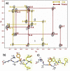
(a) Selected region of a 350 ms NOESY spectrum of [Y]6-AII (90% H2O/10% D2O). The red and green lines denote the NOE connectivities for the trans and cis isomers, respectively. Solution structures of the distinctive cis (b) and trans (c) conformers of the engineered AII analogue.
Figure 3
Figure 3
(a) Competition binding assays of [Y]6-AII analogue to AT1R, AT2R wild type, and mutants: AT1R, open circle; wild type AT2R, black circles; AT2R-Y189A, blue diamonds; AT2R-Y189N, green triangle; AT2R-F272(6.51)A, red square; AT2R-F272(6.51)H, orange triangle. Kd and Ki values are given in Supplementary Table S3. (b,c) Plots of fold selectivity values for the two AII receptor subtypes (IC50(AT1R)/IC50(AT2R)]) versus % of cis (b) and the value of Hammet substituent constants σpara (c) for the different AII analogues (see also Supplementary Table S5). (d) PC12W cells, either transduced with the Ad-AT2R or untransduced, were used for the evaluation of the AT2R agonistic effect of [Y]6-AII in the presence of either 1 nM AII or [Y]6-AII. Agonist-induced neurite outgrowth by AII or [Y]6-AII for 24 h stimulation was quantified by counting neurite-positive cell numbers in five randomly selected photos/well. The neurite outgrowth-positive cells were defined as the cells with neurite length longer than their cell diameters. This experiment was carried out in triplicate and repeated twice.
Figure 4
Figure 4
Structure of [Y]6-AII in complex with AT1R (a) and AT2R (b). [Y]6-AII is shown in yellow stick and surface, and unconserved regions in AT1R and AT2R are shown in red stick and surfaces. Red dashed lines correspond to hydrogen bonds, and green dashed lines to hydrophobic contacts. Residues depicted in blue stick were mutated for validation of the binding mode.

References

    1. Venkatakrishnan A. J.; Deupi X.; Lebon G.; Tate C. G.; Schertler G. F.; Babu M. M. (2013) Molecular signatures of G-protein-coupled receptors. Nature 494, 185–194. - PubMed
    1. Lagerstrom M. C.; Schioth H. B. (2008) Structural diversity of G protein-coupled receptors and significance for drug discovery. Nat. Rev. Drug Discovery 7, 339–357. - PubMed
    1. Katritch V.; Kufareva I.; Abagyan R. (2011) Structure based prediction of subtype-selectivity for adenosine receptor antagonists. Neuropharmacology 60, 108–115. - PMC - PubMed
    1. Tzakos A. G.; Bonvin A. M.; Troganis A.; Cordopatis P.; Amzel M. L.; Gerothanassis I. P.; van Nuland N. A. (2003) On the molecular basis of the recognition of angiotensin II (AII). NMR structure of AII in solution compared with the X-ray structure of AII bound to the mAb Fab131. Eur. J. Biochem. 270, 849–860. - PubMed
    1. Tzakos A. G.; Gerothanassis I. P.; Troganis A. N. (2004) On the structural basis of the hypertensive properties of angiotensin II: a solved mystery or a controversial issue?. Curr. Top Med. Chem. 4, 431–444. - PubMed

Publication types

MeSH terms

LinkOut - more resources