. 2014 May 1;74(9):2391-401.
doi: 10.1158/0008-5472.CAN-13-2652.

An integrated computational model of the bone microenvironment in bone-metastatic prostate cancer

Affiliations

An integrated computational model of the bone microenvironment in bone-metastatic prostate cancer

Arturo Araujo et al. Cancer Res. .

Abstract

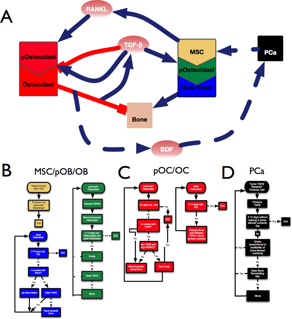
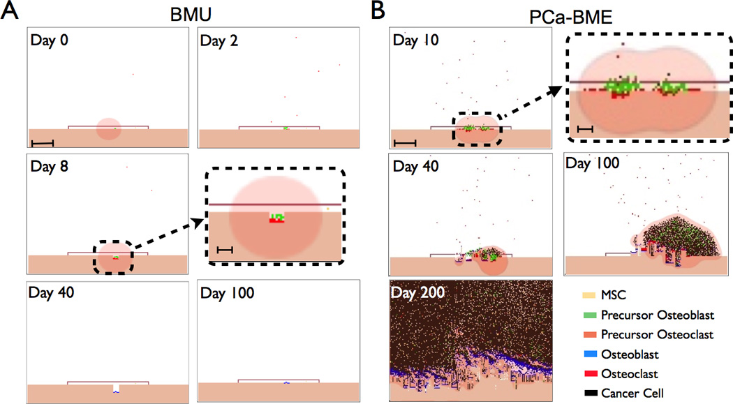
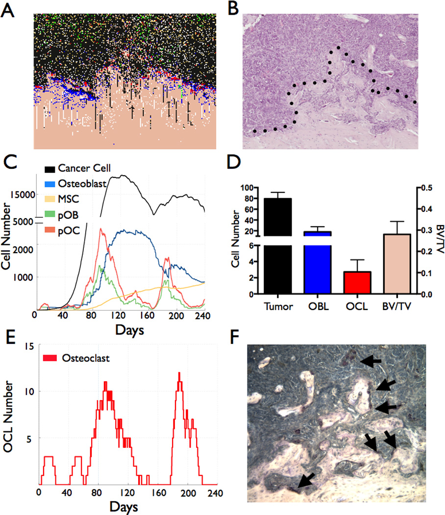
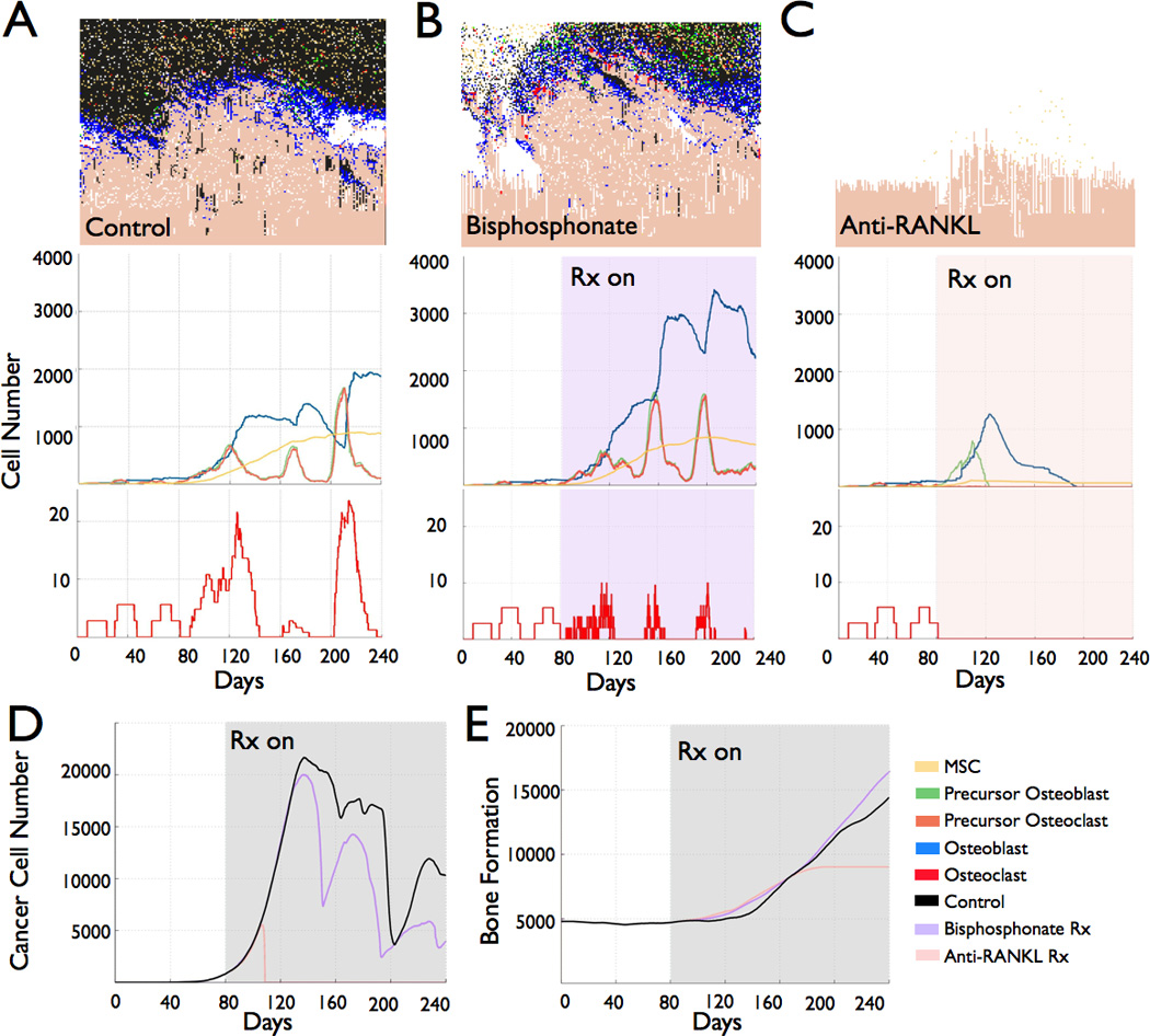
Bone metastasis will impact most men with advanced prostate cancer. The vicious cycle of bone degradation and formation driven by metastatic prostate cells in bone yields factors that drive cancer growth. Mechanistic insights into this vicious cycle have suggested new therapeutic opportunities, but complex temporal and cellular interactions in the bone microenvironment make drug development challenging. We have integrated biologic and computational approaches to generate a hybrid cellular automata model of normal bone matrix homeostasis and the prostate cancer-bone microenvironment. The model accurately reproduces the basic multicellular unit bone coupling process, such that introduction of a single prostate cancer cell yields a vicious cycle similar in cellular composition and pathophysiology to models of prostate-to-bone metastasis. Notably, the model revealed distinct phases of osteolytic and osteogenic activity, a critical role for mesenchymal stromal cells in osteogenesis, and temporal changes in cellular composition. To evaluate the robustness of the model, we assessed the effect of established bisphosphonate and anti-RANKL therapies on bone metastases. At approximately 100% efficacy, bisphosphonates inhibited cancer progression while, in contrast with clinical observations in humans, anti-RANKL therapy fully eradicated metastases. Reducing anti-RANKL yielded clinically similar results, suggesting that better targeting or dosing could improve patient survival. Our work establishes a computational model that can be tailored for rapid assessment of experimental therapies and delivery of precision medicine to patients with prostate cancer with bone metastases.


Conflict of interest statement

Disclosures of conflict of interest. None

Figures

Figure 1
Figure 1
Developing a model of the prostate tumor bone microenvironment. A, Interaction diagram showing the positive (blue) and negative (red) interactions between cell types (boxes) and factors such as TGFβ, RANKL and BDFs. B-D, Flowchart describing the sequence of steps followed by osteogenic cells (B), osteolytic cells (C) and prostate cancer cells (D) in the mathematical model.
Figure 2
Figure 2
TGFβ expression in human prostate to bone metastases. A, Patient samples (N=9) were stained for pSMAD2 (red), pan-cytokeratin (green) with nuclear contrast (DAPI). Dashed inset is magnified in panel on right. B, The intensity of pSMAD2 staining in patient samples was assessed using Definiens Tissue Studio software. C–D, TGFβ significantly enhances the migration of MSCs and MC3T3 osteoblast precursors. Representative low power objective (20×) filters illustrating MSC (C) and MC3T3 (D) migration to prostate cancer conditioned media (PAIII CM) in the presence of a TGFβ blocking antibody (1D11) or IgG control (13C4). Serum-free media (SFM) was used as a baseline control for migration. Asterisk denotes statistical significance (p<0.05).
Figure 3
Figure 3
Simulation runs from the BMU model of the bone modeling unit (BMU) and the metastatic prostate cancer bone microenvironment (PCa-BME). A, Canopy formation in response to local/systemic stimuli (Day 0). Initial osteoid degradation by retracting osteoblasts can result in the release of TGFβ that stimulates pOB expansion subsequent to asymmetric division by MSCs. Scale bar represents 250 µm. pOBs recruit pOCs in a RANKL dependent manner (Day 2). As they fuse, the pOCs become fully differentiated OCs that start resorbing bone. Inset illustrates bone resorption in the BMU. Scale bar represents 100 µm. Upon osteoclast apoptosis, pOBs differentiate into adult osteoblasts (OBs) and begin the apposition phase (Day 40). OBs rebuild bone over the course of 3 months and undergo terminal differentiation into osteocytes during the process (Day 100). B, The introduction of a TGFβ ligand and receptor expressing metastatic PCa cell perturbs BMU homeostasis (Day 0). Inset highlights tumor-bone interaction. Scale bars are 250 µm. and 100 µm respectively. The BMU canopy is compromised at Day 40 and uncontrolled bone turnover results in the enhanced recruitment of MSCs and pOCs that establishes a vicious cycle (Day 100–200).
Figure 4
Figure 4
Computational and biological model output comparisons of the prostate cancer-bone microenvironment. A–B, The computational output (A) of the prostate cancer-bone microenvironment is similar to that of an in vivo prostate cancer to bone metastasis model (B). Dashed line in B represents the tumor-pathological bone interface. C, Temporal changes in cell population in the computational model. D, Analysis of cell populations (prostate cancer cells, bone rimming cuboidal osteoblasts and TRAcP positive osteoclasts) and bone volume in biological model endpoint. E–F, Computational model outputs reveal the fluctuation of osteoclast numbers over time (E), numbers that correlate with the numbers of TRAcP osteoclasts (arrows) in similar sized fields in vivo (F).
Figure 5
Figure 5
Application of therapies to the computational model of the metastatic prostate cancer bone microenvironment. A–C, The impact of standard of care therapies on the host microenvironment cell numbers in prostate to bone metastases was assessed in simulations where no therapy was applied (Control; A), Bisphosphonates at a dosing of 4mg/5ml intravenous (Bisphosphonate; B) or RANKL targeted therapy (Anti-RANKL; C) at a dose of 120mg/1.7ml intravenous were applied at approximately 80 days post metastasis (Rx On). D–E, The impact of placebo control and standard of care therapies on tumor volume (D) and bone volume (E) over time.

References

    1. Keller ET, Brown J. Prostate cancer bone metastases promote both osteolytic and osteoblastic activity. J Cell Biochem. 2004;91:718–729. - PubMed
    1. Brown JE, Coleman RE. Denosumab in patients with cancer-a surgical strike against the osteoclast. Nature reviews Clinical oncology. 2012;9:110–118. - PubMed
    1. Bilezikian J, Raisz L, Martin T. Principles of Bone Biology. Academic Press; 2008.
    1. Bussard KM, Gay CV, Mastro AM. The bone microenvironment in metastasis; what is special about bone? Cancer Metastasis Rev. 2008;27:41–55. - PubMed
    1. Lynch CC. Matrix metalloproteinases as master regulators of the vicious cycle of bone metastasis. Bone. 2010 - PubMed

Publication types

Substances