. 2014 May 2;9(5):e94251.
doi: 10.1371/journal.pone.0094251. eCollection 2014.

Specificity of anti-tau antibodies when analyzing mice models of Alzheimer's disease: problems and solutions

Affiliations

Specificity of anti-tau antibodies when analyzing mice models of Alzheimer's disease: problems and solutions

Franck R Petry et al. PLoS One. .

Abstract

Aggregates of hyperphosphorylated tau protein are found in a group of diseases called tauopathies, which includes Alzheimer's disease. The causes and consequences of tau hyperphosphorylation are routinely investigated in laboratory animals. Mice are the models of choice as they are easily amenable to transgenic technology; consequently, their tau phosphorylation levels are frequently monitored by Western blotting using a panel of monoclonal/polyclonal anti-tau antibodies. Given that mouse secondary antibodies can recognize endogenous mouse immunoglobulins (Igs) and the possible lack of specificity with some polyclonal antibodies, non-specific signals are commonly observed. Here, we characterized the profiles of commonly used anti-tau antibodies in four different mouse models: non-transgenic mice, tau knock-out (TKO) mice, 3xTg-AD mice, and hypothermic mice, the latter a positive control for tau hyperphosphorylation. We identified 3 tau monoclonal antibody categories: type 1, characterized by high non-specificity (AT8, AT180, MC1, MC6, TG-3), type 2, demonstrating low non-specificity (AT270, CP13, CP27, Tau12, TG5), and type 3, with no non-specific signal (DA9, PHF-1, Tau1, Tau46). For polyclonal anti-tau antibodies, some displayed non-specificity (pS262, pS409) while others did not (pS199, pT205, pS396, pS404, pS422, A0024). With monoclonal antibodies, most of the interfering signal was due to endogenous Igs and could be eliminated by different techniques: i) using secondary antibodies designed to bind only non-denatured Igs, ii) preparation of a heat-stable fraction, iii) clearing Igs from the homogenates, and iv) using secondary antibodies that only bind the light chain of Igs. All of these techniques removed the non-specific signal; however, the first and the last methods were easier and more reliable. Overall, our study demonstrates a high risk of artefactual signal when performing Western blotting with routinely used anti-tau antibodies, and proposes several solutions to avoid non-specific results. We strongly recommend the use of negative (i.e., TKO) and positive (i.e., hypothermic) controls in all experiments.


Conflict of interest statement

Competing Interests: The authors have declared that no competing interests exist.

Figures

Figure 1
Figure 1. Comparison between human and mouse tau amino acidic sequence.
We reported the full-length amino acid sequence of the human and mouse tau protein containing the alternative spliced exon 2,3 and 10. We also indicated the epitopes recognized by each anti-tau primary antibodies used in this study: red: phosphorylated epitopes, black: conformational epitope, blue: human and mouse tau epitopes, green: human tau epitopes. The line between the human and mouse sequences corresponds to the sequence of amino acid recognized by the total tau primary antibody. Points correspond to a missing sequence in the species and black frames correspond to amino acid sequence differences between human and mouse species.
Figure 2
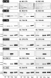
Figure 2. Analysis of tau signal with commercial monoclonal antibodies by Western blotting.
The proteins were extracted form the cortex of 3 mouse lines: control mice (WT and Hypothermic), Tau KO mice and 3xTg mice with n = 3 for each group. Proteins were extracted with SDS-PAGE and then identified with the following commercial monoclonal antibodies: A: AT8, B: AT180, C: AT270, D: Tau 12, E: Tau1 and F: Tau 46. Normal anti-mouse (1) and Mouse TrueBlot ULTRA (2) secondary antibodies were used to detect primary antibodies. The heat stable fraction was used to remove non-specificity (3). Quantifications of the blots are available in Figure S1.
Figure 3
Figure 3. Analysis of tau signal with non-commercial monoclonal antibodies by Western blotting.
The proteins were extracted form the cortex of 3 mouse lines: control mice (WT and Hypothermic), Tau KO mice and 3xTg mice with n = 3 for each group. Proteins were extracted with SDS-PAGE and then identified with the following commercial monoclonal antibodies: A: MC1, B: MC6, C: TG3, D: TG5, E: CP13, F: CP27, G: PHF-1, H: DA9. Normal anti-mouse (1) and Mouse TrueBlot ULTRA (2) secondary antibodies were used to detect primary antibodies. The heat stable fraction was used to remove non-specificity (3). Quantifications of the blots are available in Figure S2.
Figure 4
Figure 4. Effect of HS procedure on protein levels.
To determine the effect of HS procedure on protein levels, we compared the signal of Total tau (A), ERK 1/2 (B) and GAPDH (C) in control mice (WT) under 3 conditions: total sample (Total), HS fraction, and the pellet obtain after centrifugation. Data are expressed as mean ± SD, *** denotes a significant difference versus Total with P<0,001, n = 3 for each condition.
Figure 5
Figure 5. Effect of the preclearing on tau signal by Western blotting.
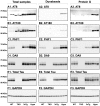
The proteins were extracted form the cortex of 3 mouse lines: control mice (WT and Hypothermic), Tau KO mice and 3xTg mice with n = 3 for each group. The supernatant obtained from tissue homogenization was processed either in the normal manner to obtain total (non-cleared) sample (1) or processed to prepare cleared samples with Dynabeads (2) or protein G (3). Protein were extracted with SDS-PAGE and then identified with the following antibodies for both total and cleared samples: A: AT8, B: AT180, C: PHF-1, D: DA9, E: total tau, F: GAPDH. Quantifications of the blots are available in Figure S3.
Figure 6
Figure 6. Analysis of tau signal with anti-mouse light chain secondary antibodies by Western blotting.
The proteins were extracted form the cortex of 3 mouse lines: control mice (WT and Hypothermic), Tau KO mice and 3xTg mice with n = 3 for each group. Proteins were extracted with SDS-PAGE and then identified with the following commercial monoclonal antibodies: A: AT8, B: AT270, C: Tau46 and D: GAPDH. We used one antibodies of each non-specificity category (AT8 for high non-specificity, AT270 for moderate non-specificity and Tau46 for absence of non-specificity). Anti-mouse light chain secondary antibodies were used to detect primary antibodies except for GAPDH. Quantifications of the blots are available in Figure S4.
Figure 7
Figure 7. Analysis of tau signal with polyclonal antibodies by Western blotting.
Proteins were extracted from the cortex of 3 mouse lines: control mice (WT and Hypothermic), Tau KO mice and 3xTg-AD mice. Proteins were separated by SDS-PAGE and then identified with the following polyclonal antibodies: A: Total Tau, B: pS199, C: pS396, D: pS404, E: pT205, F: pS422, G: pS262 and H: pS409. Normal anti-rabbit secondary antibodies were used to detect primary antibodies. The heat stable fraction was used to remove non-specificity: I: pS262 and J: pS409. Quantifications of the blots are available in Figure S5.

References

    1. Querfurth HW, LaFerla FM (2010) Alzheimer's disease. The New England journal of medicine 362: 329–344. - PubMed
    1. Selkoe DJ (2001) Alzheimer's disease: genes, proteins, and therapy. Physiological reviews 81: 741–766. - PubMed
    1. Grundke-Iqbal I, Iqbal K, Tung YC, Quinlan M, Wisniewski HM, et al. (1986) Abnormal phosphorylation of the microtubule-associated protein tau (tau) in Alzheimer cytoskeletal pathology. Proceedings of the National Academy of Sciences of the United States of America 83: 4913–4917. - PMC - PubMed
    1. Buée L, Bussière T, Buée-Scherrer V, Delacourte A, Hof PR (2000) Tau protein isoforms, phosphorylation and role in neurodegenerative disorders. Brain research Brain research reviews 33: 95–130. - PubMed
    1. Alonso AC, Zaidi T, Grundke-Iqbal I, Iqbal K (1994) Role of abnormally phosphorylated tau in the breakdown of microtubules in Alzheimer disease. Proceedings of the National Academy of Sciences of the United States of America 91: 5562–5566. - PMC - PubMed

Publication types