

The morphometry of soft tissue insertions on the tibial plateau: data acquisition and statistical shape analysis
- PMID: 24788908
- PMCID: PMC4008582
- DOI: 10.1371/journal.pone.0096515
The morphometry of soft tissue insertions on the tibial plateau: data acquisition and statistical shape analysis
Abstract
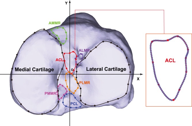
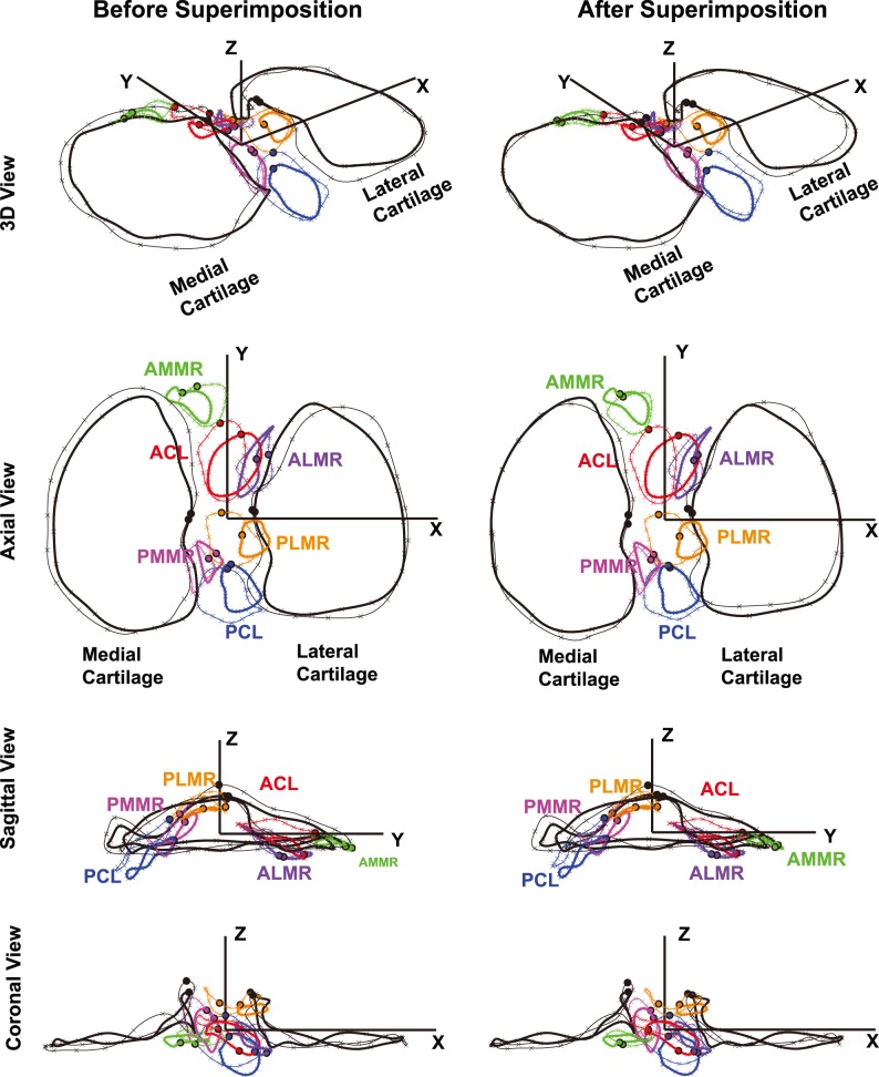
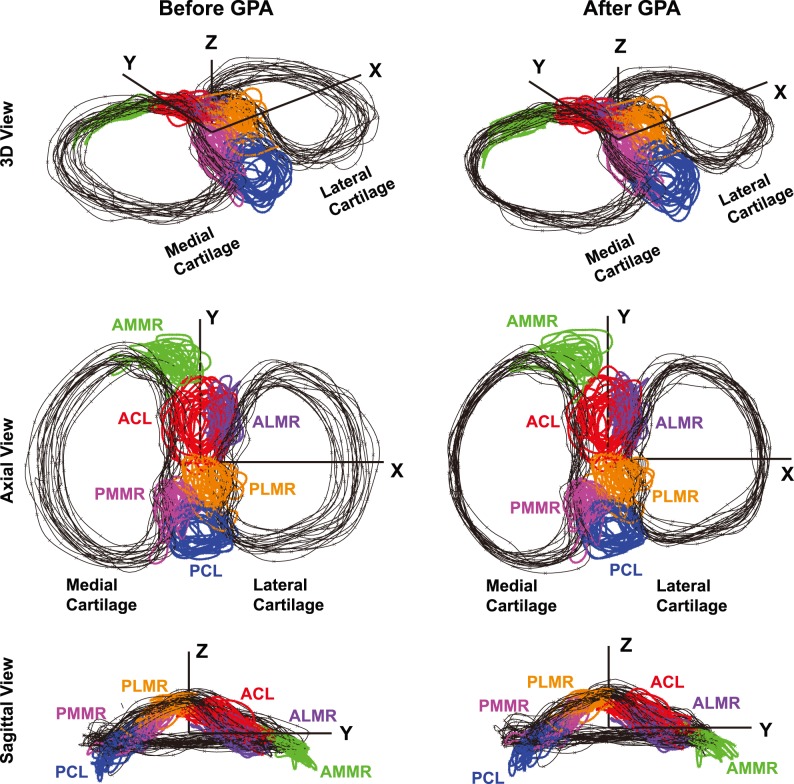
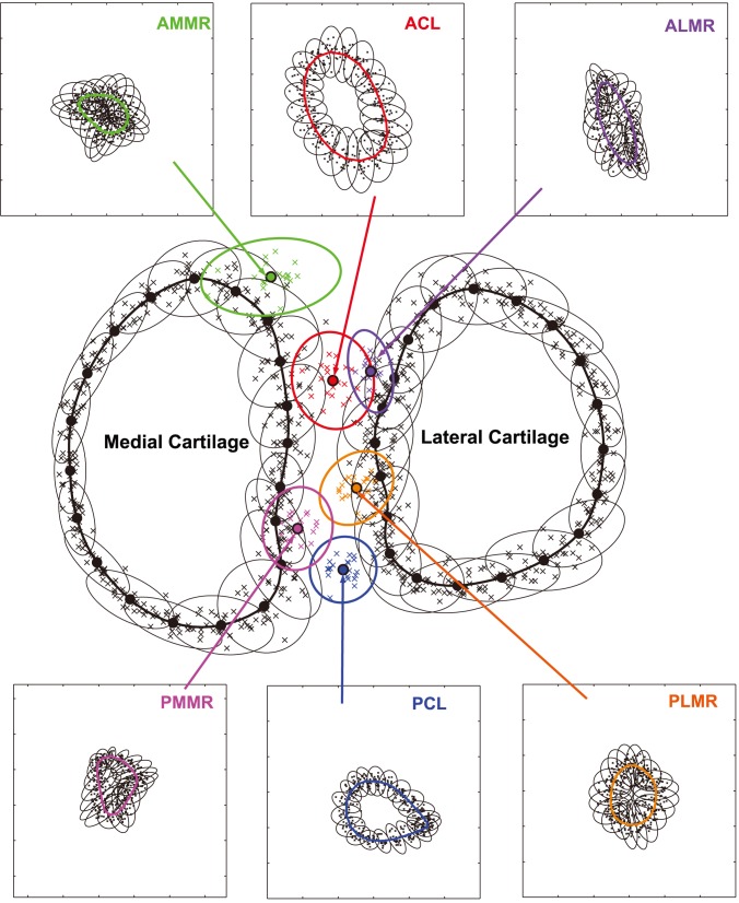
This study characterized the soft tissue insertion morphometrics on the tibial plateau and their inter-relationships as well as variabilities. The outlines of the cruciate ligament and meniscal root insertions along with the medial and lateral cartilage on 20 cadaveric tibias (10 left and 10 right knees) were digitized and co-registered with corresponding CT-based 3D bone models. Generalized Procrustes Analysis was employed in conjunction with Principal Components Analysis to first create a geometric consensus based on tibial cartilage and then determine the means and variations of insertion morphometrics including shape, size, location, and inter-relationship measures. Step-wise regression analysis was conducted in search of parsimonious models relating the morphometric measures to the tibial plateau width and depth, and basic anthropometric and gender factors. The analyses resulted in statistical morphometric representations for Procrustes-superimposed cruciate ligament and meniscus insertions, and identified only a few moderate correlations (R2: 0.37-0.49). The study provided evidence challenging the isometric scaling based on a single dimension frequently employed in related morphometric studies, and data for evaluating cruciate ligament reconstruction strategies in terms of re-creating the native anatomy and minimizing the risk of iatrogenic injury. It paved the way for future development of computer-aided personalized orthopaedic surgery applications improving the quality of care and patient safety, and biomechanical models with a better population or average representation.
Conflict of interest statement
Figures
Similar articles
-
The location of femoral and tibial tunnels in anatomic double-bundle anterior cruciate ligament reconstruction analyzed by three-dimensional computed tomography models.J Bone Joint Surg Am. 2010 Jun;92(6):1418-26. doi: 10.2106/JBJS.I.00654. J Bone Joint Surg Am. 2010. PMID: 20516317
-
Posterolateral portal tibial tunnel drilling for posterior cruciate ligament reconstruction: technique and evaluation of safety and tunnel position.Knee Surg Sports Traumatol Arthrosc. 2017 Aug;25(8):2474-2480. doi: 10.1007/s00167-015-3958-0. Epub 2015 Dec 30. Knee Surg Sports Traumatol Arthrosc. 2017. PMID: 26718637
-
Anterior cruciate ligament bundle insertions vary between ACL-rupture and non-injured knees.Knee Surg Sports Traumatol Arthrosc. 2021 Apr;29(4):1164-1172. doi: 10.1007/s00167-020-06122-1. Epub 2020 Jul 1. Knee Surg Sports Traumatol Arthrosc. 2021. PMID: 32613337
-
Variations in Knee Kinematics After ACL Injury and After Reconstruction Are Correlated With Bone Shape Differences.Clin Orthop Relat Res. 2017 Oct;475(10):2427-2435. doi: 10.1007/s11999-017-5368-8. Clin Orthop Relat Res. 2017. PMID: 28451863 Free PMC article.
-
Anatomical study of the femoral and tibial insertions of the anterolateral and posteromedial bundles of human posterior cruciate ligament.Knee Surg Sports Traumatol Arthrosc. 2006 Nov;14(11):1055-9. doi: 10.1007/s00167-006-0192-9. Epub 2006 Aug 4. Knee Surg Sports Traumatol Arthrosc. 2006. PMID: 16897069
Cited by
-
The accuracy of a newly developed guide system in medial meniscus posterior root repair: a comparison between two aiming guides.Knee Surg Relat Res. 2019 Aug 7;31(1):7. doi: 10.1186/s43019-019-0007-1. Knee Surg Relat Res. 2019. PMID: 32660577 Free PMC article.
-
Morphometric Analysis of the Tibial Attachment Shape of the Anterior Cruciate Ligament and Its Relationship With the Location of the Anterior Horn of the Lateral Meniscus.Am J Sports Med. 2024 Mar;52(3):682-690. doi: 10.1177/03635465231219978. Epub 2024 Jan 29. Am J Sports Med. 2024. PMID: 38284162 Free PMC article.
-
Systematic Review of Cadaveric Studies on Anterior Cruciate Ligament Anatomy Focusing on the Mid-substance Insertion and Fan-like Extension Fibers.Indian J Orthop. 2022 Jul 18;56(9):1525-1532. doi: 10.1007/s43465-022-00695-4. eCollection 2022 Sep. Indian J Orthop. 2022. PMID: 36052387 Free PMC article. Review.
-
Estimation of footprints of the canine stifle ligaments using deformable shape templates of bones.Sci Rep. 2024 Feb 26;14(1):4639. doi: 10.1038/s41598-024-55116-3. Sci Rep. 2024. PMID: 38409316 Free PMC article.
-
Significance of the broad non-bony attachments of the anterior cruciate ligament on the tibial side.Sci Rep. 2022 Apr 27;12(1):6844. doi: 10.1038/s41598-022-10806-8. Sci Rep. 2022. PMID: 35477722 Free PMC article.
References
-
- Plaweski S, Petek D, Saragaglia D (2011) Morphometric analysis and functional correlation of tibial and femoral footprints in anatomical and single bundle reconstructions of the anterior cruciate ligament of the knee. Orthop Traumatol Surg Res 97: S75–79. - PubMed
-
- Zantop T, Diermann N, Schumacher T, Schanz S, Fu FH, et al. (2008) Anatomical and nonanatomical double-bundle anterior cruciate ligament reconstruction: importance of femoral tunnel location on knee kinematics. Am J Sports Med 36: 678–685. - PubMed
-
- Simmons R, Howell SM, Hull ML (2003) Effect of the angle of the femoral and tibial tunnels in the coronal plane and incremental excision of the posterior cruciate ligament on tension of an anterior cruciate ligament graft: an in vitro study. J Bone Joint Surg Am 85-A: 1018–1029. - PubMed
-
- Iriuchishima T, Tajima G, Ingham SJ, Shirakura K, Fu FH (2012) PCL to graft impingement pressure after anatomical or non-anatomical single-bundle ACL reconstruction. Knee Surg Sports Traumatol Arthrosc 20: 964–969. - PubMed
-
- Galloway MT, Grood ES, Mehalik JN, Levy M, Saddler SC, et al. (1996) Posterior cruciate ligament reconstruction. An in vitro study of femoral and tibial graft placement. Am J Sports Med 24: 437–445. - PubMed
MeSH terms
Grants and funding
LinkOut - more resources
Full Text Sources
Other Literature Sources
