. 2014 May 1;9(5):e94989.
doi: 10.1371/journal.pone.0094989. eCollection 2014.

Disrupted bone remodeling leads to cochlear overgrowth and hearing loss in a mouse model of fibrous dysplasia

Affiliations

Disrupted bone remodeling leads to cochlear overgrowth and hearing loss in a mouse model of fibrous dysplasia

Omar Akil et al. PLoS One. .

Abstract

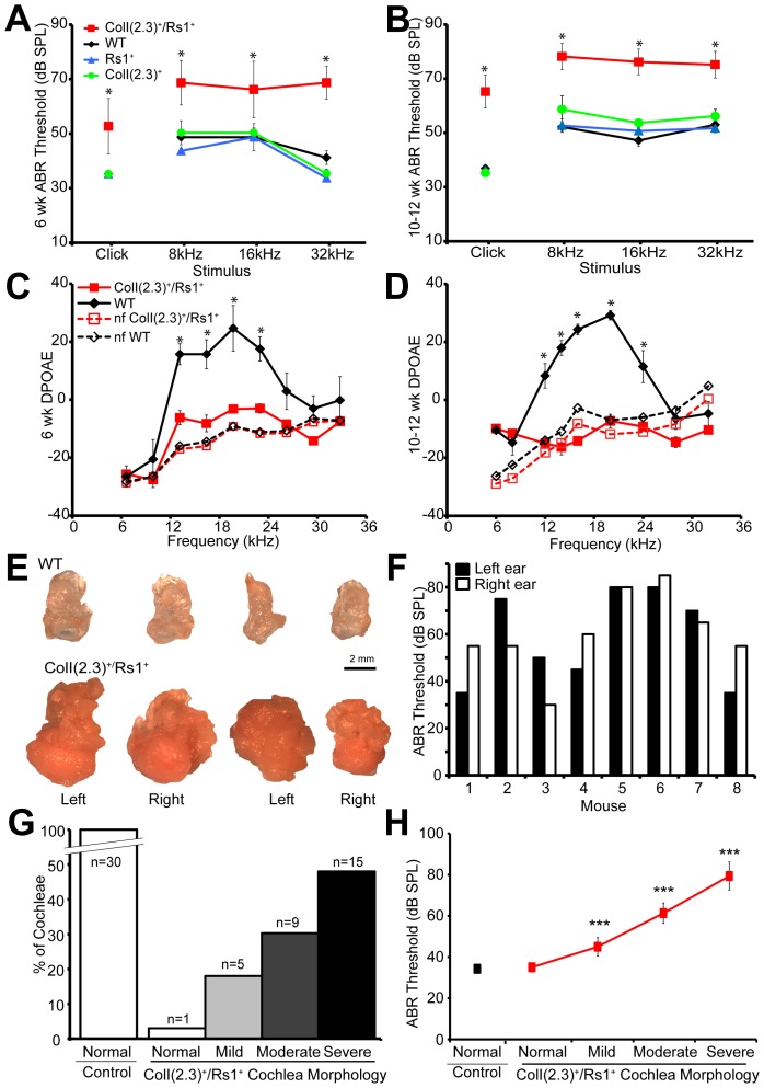
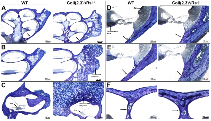
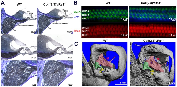
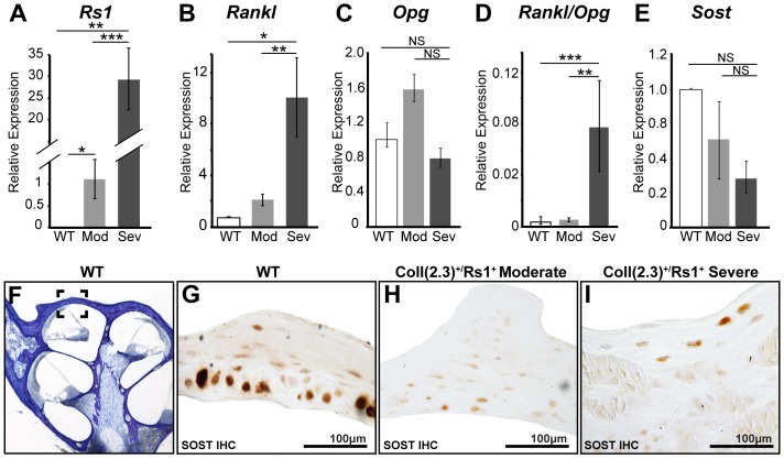
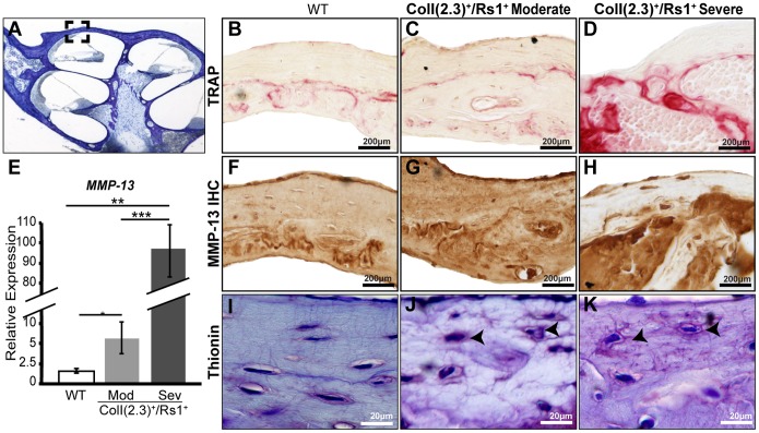
Normal hearing requires exquisite cooperation between bony and sensorineural structures within the cochlea. For example, the inner ear secretes proteins such as osteoprotegrin (OPG) that can prevent cochlear bone remodeling. Accordingly, diseases that affect bone regulation can also result in hearing loss. Patients with fibrous dysplasia develop trabecular bone overgrowth resulting in hearing loss if the lesions affect the temporal bones. Unfortunately, the mechanisms responsible for this hearing loss, which could be sensorineural and/or conductive, remain unclear. In this study, we used a unique transgenic mouse model of increased Gs G-protein coupled receptor (GPCR) signaling induced by expression of an engineered receptor, Rs1, in osteoblastic cells. These ColI(2.3)+/Rs1+ mice showed dramatic bone lesions that histologically and radiologically resembled fibrous dysplasia. We found that ColI(2.3)+/Rs1+ mice showed progressive and severe conductive hearing loss. Ossicular chain impingement increased with the size and number of dysplastic lesions. While sensorineural structures were unaffected, ColI(2.3)+/Rs1+ cochleae had abnormally high osteoclast activity, together with elevated tartrate resistant acid phosphatase (TRAP) activity and receptor activator of nuclear factor kappa-B ligand (Rankl) mRNA expression. ColI(2.3)+/Rs1+ cochleae also showed decreased expression of Sclerostin (Sost), an antagonist of the Wnt signaling pathway that normally increases bone formation. The osteocyte canalicular networks of ColI(2.3)+/Rs1+ cochleae were disrupted and showed abnormal osteocyte morphology. The osteocytes in the ColI(2.3)+/Rs1+ cochleae showed increased expression of matrix metalloproteinase 13 (MMP-13) and TRAP, both of which can support osteocyte-mediated peri-lacunar remodeling. Thus, while the ossicular chain impingement is sufficient to account for the progressive hearing loss in fibrous dysplasia, the deregulation of bone remodeling extends to the cochlea as well. Our findings suggest that factors regulating bone remodeling, including peri-lacunar remodeling by osteocytes, may be useful targets for treating the bony overgrowths and hearing changes of fibrous dysplasia and other bony pathologies.


Conflict of interest statement

Competing Interests: The authors have declared that no competing interests exist.

Figures

Figure 1
Figure 1. Progressive hearing loss observed in ColI(2.3)+/Rs1+ mice with fibrous dysplasia-like lesions correlates to the severity of dysplastic cochlear lesions.
(A, B) ABR thresholds (decibels of sound pressure level, dBSPL) were measured in 6-week-old and 10–12-week-old ColI(2.3)+/Rs1+ (mutant), WT, and ColI(2.3)−/Rs1+ and ColI(2.3)+/Rs1 (single transgenic) mice. ColI(2.3)+/Rs1+ mice showed higher ABR thresholds at. Click stimulus and at 8, 16, and 32 kHz stimuli when compared to WT and single transgenic mice at both 6- and 10–12-week-old time points. Male and female mice were analyzed together. (C) DPOAEs were also measured at 6-weeks and 10–12-weeks. WT and ColI(2.3)+/Rs1+ mice and compared to WT and ColI(2.3)+/Rs1+ background noise floor (nf) measurements of all experimental noise sources used. ColI(2.3)+/Rs1+ mutant DPOAEs at 6, 12, 18, 24, 32 kHz frequencies showed levels similar to nf controls compared to normal WT DPOAEs at 6 weeks of age. Male and female mice were analyzed together. (D) Increased differences between WT and ColI(2.3)+/Rs1+ DPOAE recordings were observed in 10–12 week-old mice. *, p<0.05. (E) Images of dissected cochleae from two representative sets of ColI(2.3)+/Rs1+ and WT 12-week-old mice showed gross abnormalities caused by fibrous lesion growth compared to WT cochlea. Scale bar = 2 mm. (F) Click ABR thresholds were measured in the left and right ears of 12-week mice (n = 8), with highly variable differences observed in the same mouse. Male and female mice were analyzed together. (G) A rank-order histological grading system (see methods for criteria) was used to group the severity of ColI(2.3)+/Rs1+ cochlear lesions into normal, mild, moderate, and severe lesions from 30 ColI(2.3)+/Rs1+ cochleae. When compared to 30 normal WT cochleae which all had normal morphology, chi-squared analysis showed p<0.0001. (H) The categorized lesions from 15 control (30 cochleae) and 15 ColI(2.3)+/Rs1+ mice (30 cochleae) were compared to measured ABR thresholds. N = 1 cochlea that appeared normal; 5 cochleae with mild lesions; 9 cochleae with moderate lesions; and 15 cochlea with severe lesions, as in (G). Male and female mice were analyzed together. ***, p<0.0001 when compared to control ABR threshold.
Figure 2
Figure 2. Irregular lesions in ColI(2.3)+/Rs1+ cochlea involve the apex and the labyrinth causing thickening of the otic capsule.
(A–C) Cochlea from 12-week-old WT and ColI(2.3)+/Rs1+ mice were stained for toluidine blue and examined histologically. Mixed boney fibrous lesions were often observed surrounding ColI(2.3)+/Rs1+ cochlea compared to WT controls. ColI(2.3)+/Rs1+ cochlea had multiple fibrous boney overgrowths of the vestibular bone compared to normal WT morphology. Scale bar, 150 µm. (D–F) The walls of ColI(2.3)+/Rs1+ cochleae showed significant thickening in comparison to WT cochleae, possibly due to the overall thickening of the surrounding otic capsule. The stria vascularis (SV) in the ColI(2.3)+/Rs1+ mice appear normal. Scale bar, 100 µm.
Figure 3
Figure 3. Sensorineural structures of ColI(2.3)+/Rs1+ mice are normal despite the bony overgrowth affecting the ossicular chain.
(A) Histological analysis of the sensorineural structures of 12-week-old WT and ColI(2.3)+/Rs1+ revealed no gross abnormalities in the organ of Corti (OC), tunnel of Corti (TC), inner hair cells (IHC), or outer hair cells (OHC). (B) Whole mount immunofluorescence preparations of the outer hair cells revealed no visible differences in hair cell structure or number in ColI(2.3)+/Rs1+ in comparison to WT structures. (C) Micro computed x-ray tomography of the temporal bones was examined. A region of interest including the middle ear was selected in WT and ColI(2.3)+/Rs1+ at 12-week-old mice. ColI(2.3)+/Rs1+ temporal bones showed significant bony overgrowth of the middle ear compared to WT. The ossicles are structurally identifiable and are affected in the ColI(2.3)+/Rs1+ mice [malleus (M), incus (I), and stapes (S)].
Figure 4
Figure 4. Markers of osteoclast-mediated bone remodeling is reactivated in ColI(2.3)+/Rs1+ cochlea.
Markers of bone remodeling were assessed by immunohistochemistry on dissected 12-week-old male WT and ColI(2.3)+/Rs1+ cochlea with moderate (Mod) and severe (Sev) fibrous lesions using quantitative PCR. (A) Rs1 transgene expression was absent in WT controls and elevated in moderate (**, p = 0.01) and elevated 30-fold in severe ColI(2.3)+/Rs1+ cochlea (***, p<0.001). (B) Rankl, a marker for osteoclast differentiation and activity was significantly increased in ColI(2.3)+/Rs1+ severe cochlea when compared to WT and moderate ColI(2.3)+/Rs1+ cochlea (*, p<0.05; **, p<0.01). No statistically significant differences were observed in moderate ColI(2.3)+/Rs1+ cochlea compared to WT (p = 0.63). (C) There were also no statistically significant changes observed in Osteoprotegrin expression in moderate and severe ColI(2.3) +/Rs1+ cochlea compared to WT cochlea (p = 0.29, and p = 0.645 respectively). (D) The ratios of Rankl and osteoclast inhibitory factor osteoprotegrin (Opg) were significantly increased in ColI(2.3)+/Rs1+ severe cochlea compared to moderate ColI(2.3)+/Rs1+ (**, p<0.01) and WT cochlea (***, p<0.005) indicating that osteoclast mediated remodeling is increased in ColI(2.3)+/Rs1+ severe cochlea. (E) No statistically significant changes were observed in Sost expression in ColI(2.3)+/Rs1+ moderate and severe cochlea compared to WT expression (p = 0.73 and p = 0.43 respectively). Error bars are mean +/− SEM of triplicate measurements, n = 5 WT and 5 each of ColI(2.3)+/Rs1+ moderate and severe cochleae. (F) A specific area of the otic capsule (indicated by box) was examined immunohistochemical analysis. (G–I) Immunohistochemistry for SOST showed visibly significant decreases in expression in moderate and severe ColI(2.3)+/Rs1+ cochlea compared to WT cochlea.
Figure 5
Figure 5. Abnormal peri-lacunar remodeling in cochlear fibrous dysplasia.
(A) 12-week-old cochlea from male WT and ColI(2.3)+/Rs1+ cochleae were examined for markers of peri-lacunar remodeling in a specific area of the otic capsule. (B–D) TRAP, a marker for osteoclast activity, was elevated in moderate and severe ColI(2.3)+/Rs1+ otic capsules compared to the low levels observed in WT controls. (E) The expression of osteocyte secreted remodeling factor, MMP-13 was significantly increased 7-fold in moderate (**, p<0.005) and 100 fold in severe ColI(2.3)+/Rs1+ cochleae (***, p<1×10−05) compared to WT cochleae. (n = 6 male WT, n = 5 male ColI(2.3)+/Rs1+ moderate, and n = 5 male ColI(2.3)+/Rs1+ severe cochlea). Error bars are mean +/− SEM of triplicate measurements. (F–H) Immunohistochemistry showed that MMP-13 expression was confined to the otic capsule in WT cochleae, but the domain of expression is increased in ColI(2.3)+/Rs1+ moderate and severe cochlea. (I–K) Thionin staining of the canalicular network was examined in WT and ColI(2.3)+/Rs1+ moderate and severe cochleae. The canalicular network in WT cochlea shows normal elliptical osteocyte morphology and connectivity. ColI(2.3)+/Rs1+ moderate and severe cochleae show an abnormal rounded osteocyte morphologies with disrupted and disorganized canalicular networks of varying severity (n = 5 males per genotype).

Similar articles

Cited by

References

    1. Raggatt LJ, Partridge NC (2010) Cellular and molecular mechanisms of bone remodeling. The Journal of biological chemistry 285: 25103–25108. - PMC - PubMed
    1. Feng X, McDonald JM (2011) Disorders of bone remodeling. Annual review of pathology 6: 121–145. - PMC - PubMed
    1. Zehnder AF, Kristiansen AG, Adams JC, Kujawa SG, Merchant SN, et al. (2006) Osteoprotegrin knockout mice demonstrate abnormal remodeling of the otic capsule and progressive hearing loss. The Laryngoscope 116: 201–206. - PMC - PubMed
    1. Lyford-Pike S, Hoover-Fong J, Tunkel DE (2012) Otolaryngologic manifestations of skeletal dysplasias in children. Otolaryngologic clinics of North America 45: 579–598, vii. - PubMed
    1. Chang JL, Brauer DS, Johnson J, Chen CG, Akil O, et al. (2010) Tissue-specific calibration of extracellular matrix material properties by transforming growth factor-beta and Runx2 in bone is required for hearing. EMBO reports 11: 765–771. - PMC - PubMed

Publication types

LinkOut - more resources