. 2014 May 2;9(5):e96092.
doi: 10.1371/journal.pone.0096092. eCollection 2014.

Coordinated regulation of nuclear receptor CAR by CCRP/DNAJC7, HSP70 and the ubiquitin-proteasome system

Affiliations

Coordinated regulation of nuclear receptor CAR by CCRP/DNAJC7, HSP70 and the ubiquitin-proteasome system

Yoav E Timsit et al. PLoS One. .

Abstract

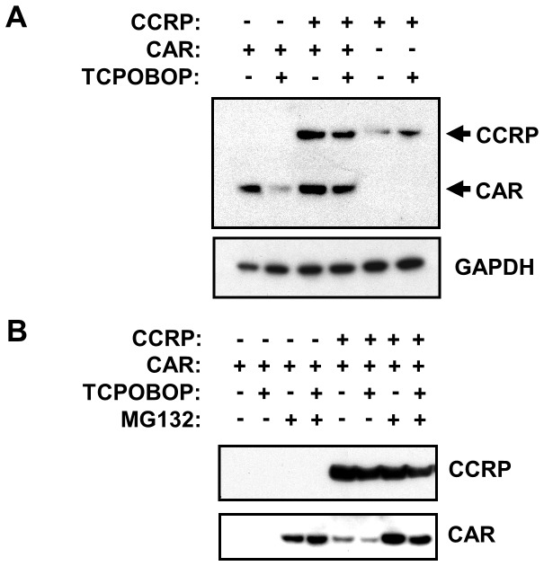
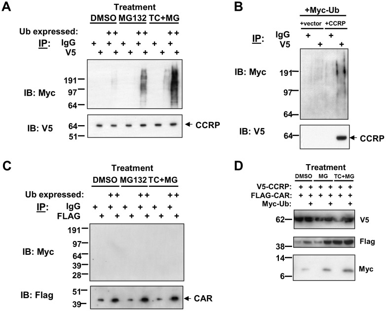
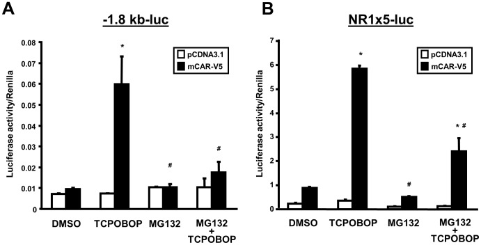
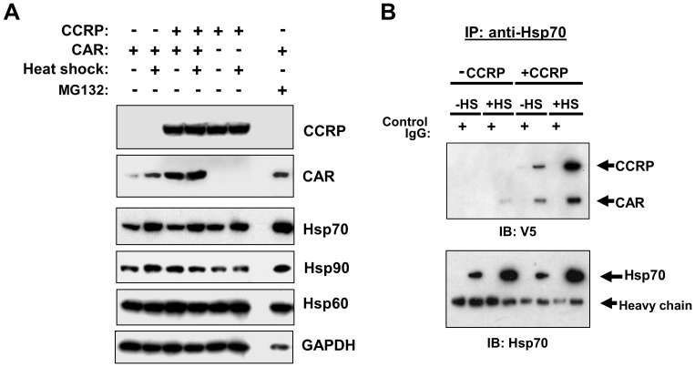
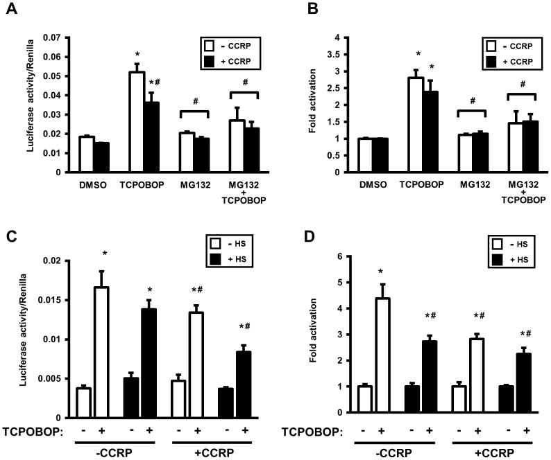
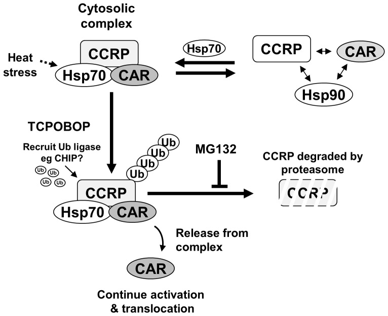
The constitutive active/androstane receptor (CAR) plays an important role as a coordinate transcription factor in the regulation of various hepatic metabolic pathways for chemicals such as drugs, glucose, fatty acids, bilirubin, and bile acids. Currently, it is known that in its inactive state, CAR is retained in the cytoplasm in a protein complex with HSP90 and the tetratricopeptide repeat protein cytosoplasmic CAR retention protein (CCRP). Upon activation by phenobarbital (PB) or the PB-like inducer 1,4-bis[2-(3,5-dichloropyridyloxy)]-benzene (TCPOBOP), CAR translocates into the nucleus. We have identified two new components to the cytoplasmic regulation of CAR: ubiquitin-dependent degradation of CCRP and protein-protein interaction with HSP70. Treatment with the proteasome inhibitor MG132 (5 µM) causes CAR to accumulate in the cytoplasm of transfected HepG2 cells. In the presence of MG132, TCPOBOP increases CCRP ubiquitination in HepG2 cells co-expressing CAR, while CAR ubiquitination was not detected. MG132 treatment of HepG2 also attenuated of TCPOBOP-induced CAR transcriptional activation on reporter constructs which contain CAR-binding DNA elements derived from the human CYP2B6 gene. The elevation of cytoplasmic CAR protein with MG132 correlated with an increase of HSP70, and to a lesser extent HSP60. Both CCRP and CAR were found to interact with endogenous HSP70 in HepG2 cells by immunoprecipitation analysis. Induction of HSP70 levels by heat shock also increased cytoplasmic CAR levels, similar to the effect of MG132. Lastly, heat shock attenuated TCPOBOP-induced CAR transcriptional activation, also similar to the effect of MG132. Collectively, these data suggest that ubiquitin-proteasomal regulation of CCRP and HSP70 are important contributors to the regulation of cytoplasmic CAR levels, and hence the ability of CAR to respond to PB or PB-like inducers.


Conflict of interest statement

Competing Interests: The authors have declared that no competing interests exist.

Figures

Figure 1
Figure 1. Stabilization of CAR in the cytoplasm.
(A) HepG2 cells were transfected with V5-tagged mouse CAR (pcDNA3.1/V5-His-mCAR-V5, 3 µg) and mouse CCRP (pcDNA3.1/V5-His-mCCRP, 0.3 µg), and 18 hours later transfected cells were treated with DMSO (0.1% v/v) or TCPOBOP (250 nM) for 24 hr. Cells were then harvested and cytosolic extracts prepared and subjected to immunoblotting analysis with anti-V5 antibodies. (B) HepG2 cells were transfected with V5-tagged CCRP (pcDNA3.1-mCCRP-V5, 0.3 µg) or empty vector (pcDNA3.1-V5) combined with FLAG-tagged mCAR (pCR3-mCAR-FLAG, 3 µg).18 hours after transfection, cells were treated with DMSO (0.1% v/v), TCPOBOP (250 nM in 0.1% DMSO, final concentrations), MG132 (5 µM in 0.1% DMSO, final concentrations), or TCPOBOP combined with MG132 for 24 hr. Cells were then harvested and cytosolic extracts prepared and subjected to immunoblotting analysis with anti-V5 antibodies (upper panel) and with anti-FLAG antibodies (lower panel). Results shown are representative of three independent experiments.
Figure 2
Figure 2. CCRP is ubiquitinated upon TCPOBOP treatment.
Various combinations of the following plasmids were transfected into HepG2 cells 18(cell density 5×10⁶ cells/10 cm plate): pcDNA3.1/V5-His-mCCRP (3 µg), pCR3-mCAR-FLAG (3 µg), and pCW7-Myc-Ub (3 µg). 24 hr after transfection, cells were treatment with DMSO (0.1% v/v), MG132 (5 µM in 0.1 1% DMSO, final concentrations) or MG132 (5 µM) in combination with TCPOBOP (250 nM dissolved in 0.1% DMSO, final concentrations). At the end of treatment, cells were harvested, cytosolic extracts prepared, and immunoprecipitation analysis was performed to detect ubiquitinated CCRP and CAR. Results shown are representative of three independent experiments. (A) Cells were transfected pcDNA3.1/V5-His-mCCRP (3 µg) and pCR3-mCAR-FLAG (3 µg) with or without pCW7-Myc-Ub (3 µg). Immunoprecipitation using anti-V5 antibodies or anti-IgG (as control) was performed, followed by immunoblotting analysis using anti-V5 and anti-Myc antibodies. (B) As a control for detecting ubiquitinated CCRP, extracts derived from cells cotransfected with empty vector or pcDNA3.1/V5-His-mCCRP and pCW7-Myv-Ub were subjected to immunoprecipation then immunoblotting analysis in (A). (C) Extracts were also subjected to imunoprecipitation analysis using anti-FLAG antibodies or anti-IgG (as control), followed by immunoblotting analysis using anti-FLAG and anti-Myc antibodies. (D) Immunblotting results of extracts used in the immunoprecipitation experiments, using anti-V5, anti-FLAG, and anti-Myc antibodies.
Figure 3
Figure 3. Proteasomal inhibition attenuates TCPOBOP-induced CAR transcriptional activation in HepG2 cells.
Cells were cotransfected 18/V5-His-mCAR or empty vector combined with either the luciferase reporter plasmid containing −1.8 kb upstream region of the CYP2B6 gene promoter (−1.8-kb-luc) (A) or the (NR1)5-tk-luciferase [(NR1)5-tk-luc] reporter plasmid (B). The Renilla construct (phRL-tk) was also co-transfected as a normalization control. Cells were then treated with DMSO (0.1% v/v), TCPOBOP (250 nM dissolved in 0.1% DMSO, final concentrations), MG132 (5 µM in 0.1 1% DMSO, final concentrations), or MG132 plus TCPOBOP, after which cells were harvested and luciferase activity measured. Luciferase activity of each sample was normalized to Renilla activity, and expressed as means ± SD of triplicate determinations. Results shown are representative of three independent experiments. *Significantly different (p<0.0001) compared to DMSO (+mCAR-V5); #significantly different (p<0.0001) compared to TCPOPOP (+mCAR-V5), based on one-way ANOVA with post-hoc Tukey multiple comparisons test.
Figure 4
Figure 4. Increased levels of cytoplasmic CAR by proteasomal inhibition correlates with an increase in HSP70 levels.
Results shown are representative of three independent experiments. (A) HepG2 cells were transfected with V5-tagged mouse CAR (pcDNA3.1/V5-His-mCAR, 3 µg) and mouse CCRP (pcDNA3.1/V5-His-mCCRP, 0.3 µg), and 18 hours later transfected cells were treated for 24 hr with DMSO (0.1% v/v), TCPOBOP (250 nM in 0.1% DMSO, final concentrations), MG132 (5 µM in 0.1% DMSO, final concentrations), or TCPOBOP combined with MG132. Cells were then harvested and cytosolic extracts prepared and subjected to immunoblotting analysis with antibodies against the indicated proteins. (B) Extracts analyzed in (A) were subjected to immunoprecipitation analysis using anti-V5 or anti-IgG (as control) antibodies was performed, followed by immunoblotting analysis using anti-V5 (top panel), anti-HSP70 (middle panel), and anti-HSP90 (bottom panel) antibodies.
Figure 5
Figure 5. Thermal stress increases cytoplasmic CAR in an HSP70-dependent manner.
Results shown are representative of three independent experiments. (A) HepG2 cells were cotransfected with V5-tagged CCRP (pcDNA3.1/V5-His-mCCRP, 0.3 µg) or empty vector and FLAG-tagged mCAR (pCR3-mCAR-FLAG, 3 µg) or empty vector. 18 hr after transfection, cells were incubated for 1 hr at 42°C or 37°C, followed by treatment at 37°C with DMSO (0.1% DMSO v/v) or MG132 (5 µM in 0.1%DMSO, final concentrations). Cells were then harvested and cytosolic extracts prepared and subjected to immunoblotting analysis with antibodies against the indicated proteins. (B) HepG2 cells were transfected with V5-tagged mouse CAR (pcDNA3.1/V5-His-mCAR, 3 µg) and mouse CCRP (pcDNA3.1/V5-His-mCCRP, 0.3 µg). 18 hr after transfection, cells were incubated for 1 hr at 42°C or 37°C, followed by treatment at 37°C with DMSO for 24 hr. Cell extracts were prepared and then subjected to immunoprecipitation analysis using anti-V5 or anti-IgG (as control) antibodies, followed by immunoblotting analysis using anti-V5 (top panel) and anti-HSP70 (bottom panel) antibodies.
Figure 6
Figure 6. Thermal stress attenuates TCPOBOP-induced CAR transcriptional activation in HepG2 cells, similar to the effect of proteasomal inhibition.
Luciferase activity of each sample was normalized to Renilla activity, and expressed as means ± SD of triplicate determinations. Shown is representative of three independent experiments. (A and B) Experiments were performed as described in Fig. 3, however empty vector or pcDNA3.1/V5-His-mCCRP was cotransfected with pcDNA3.1/V5-His-mCAR, -1.8-kb-luc, and phRL-tk (as normalization control). After transfection, cells were treated for 24 hr with DMSO (0.1% v/v), TCPOBOP (250 nM dissolved in 0.1% DMSO, final concentrations), MG132 (5 µM in 0.1% DMSO, final concentrations), or MG132 plus TCPOBOP, after which cells were harvested and luciferase activity measured. For fold-change determinations (B), each group was normalized to its corresponding DMSO-treated control (set as 1). Statistics are based on one-way ANOVA with post-hoc Tukey multiple comparisons test. *Significantly different (p<0.0001) compared to DMSO; and #significantly different (p<0.002) compared to TCPOPOP without CCRP overexpression. (C and D) Experiments and data analysis were performed as in A and B, with an incubation step at 42°C for 1 hr for heat-shock-designated cells performed prior to 24 hr treatment with DMSO (0.1% v/v) or TCPOBOP (250 nM in 0.1% DMSO, final concentrations). For fold-change determinations (D), each group was normalized to its corresponding DMSO-treated control (set as 1). Statistics are based on one-way ANOVA with post-hoc Tukey multiple comparisons test. *Significantly different (p<0.0001) compared to corresponding DMSO control treatment; #significantly different (p<0.001) to TCPOPOP alone (without both CCRP overexpression and heat shock).
Figure 7
Figure 7. Proposed model for the regulation of CAR by the ubiquitin-proteasome system.
An equilibrium between a stable CAR cytoplasmic complex (at top left) and a weaker associated complex exists (at top right), and HSP70 functions to favor the stable cytoplasmic complex. Activator treatment signals for ubiquitination of CCRP, resulting in its degradation by the proteasome. This results in the release of CAR from the cytosolic complex, followed by translocation into the nucleus.

References

    1. Timsit YE, Negishi M (2007) CAR and PXR: the xenobiotic-sensing receptors. Steroids 72: 231–246. - PMC - PubMed
    1. Ueda A, Hamadeh HK, Webb HK, Yamamoto Y, Sueyoshi T, et al. (2002) Diverse roles of the nuclear orphan receptor CAR in regulating hepatic genes in response to phenobarbital. Mol Pharmacol 61: 1–6. - PubMed
    1. Wei P, Zhang J, Egan-Hafley M, Liang S, Moore DD (2000) The nuclear receptor CAR mediates specific xenobiotic induction of drug metabolism. Nature 407: 920–923. - PubMed
    1. Honkakoski P, Moore R, Gynther J, Negishi M (1996) Characterization of phenobarbital-inducible mouse Cyp2b10 gene transcription in primary hepatocytes. J Biol Chem 271: 9746–9753. - PubMed
    1. Honkakoski P, Negishi M (1998) Regulatory DNA elements of phenobarbital-responsive cytochrome P450 CYP2B genes. J Biochem Mol Toxicol 12: 3–9. - PubMed

Publication types

MeSH terms