. 2014 Nov;19(6):877-86.
doi: 10.1007/s12192-014-0511-x. Epub 2014 May 1.

Interaction of heat shock protein 70 with membranes depends on the lipid environment

Affiliations

Interaction of heat shock protein 70 with membranes depends on the lipid environment

Gabrielle Armijo et al. Cell Stress Chaperones. 2014 Nov.

Abstract

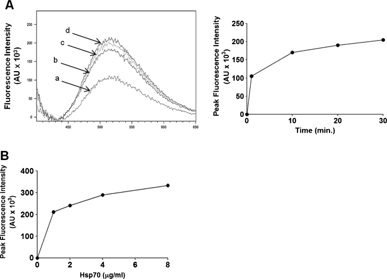
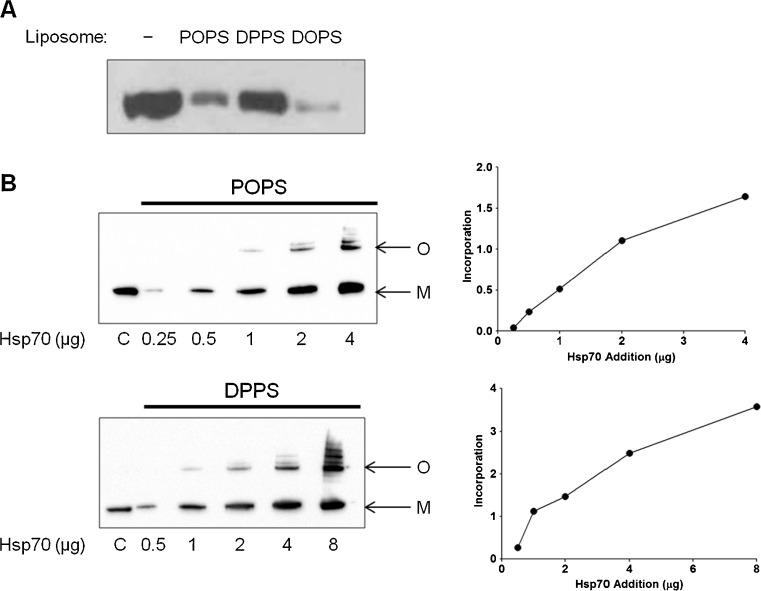
Heat shock proteins (hsp) are well recognized for their protein folding activity. Additionally, hsp expression is enhanced during stress conditions to preserve cellular homeostasis. Hsp are also detected outside cells, released by an active mechanism independent of cell death. Extracellular hsp appear to act as signaling molecules as part of a systemic response to stress. Extracellular hsp do not contain a consensus signal for their secretion via the classical ER-Golgi compartment. Therefore, they are likely exported by an alternative mechanism requiring translocation across the plasma membrane. Since Hsp70, the major inducible hsp, has been detected on surface of stressed cells, we propose that membrane interaction is the first step in the export process. The question that emerges is how does this charged cytosolic protein interact with lipid membranes? Prior studies have shown that Hsp70 formed ion conductance pathways within artificial lipid bilayers. These early observations have been extended herewith using a liposome insertion assay. We showed that Hsp70 selectively interacted with negatively charged phospholipids, particularly phosphatidyl serine (PS), within liposomes, which was followed by insertion into the lipid bilayer, forming high-molecular weight oligomers. Hsp70 displayed a preference for less fluid lipid environments and the region embedded into the lipid membrane was mapped toward the C-terminus end of the molecule. The results from our studies provide evidence of an unexpected ability of a large, charged protein to become inserted into a lipid membrane. This observation provides a new paradigm for the interaction of proteins with lipid environments. In addition, it may explain the export mechanism of an increasing number of proteins that lack the consensus secretory signals.


Figures

Fig. 1
Fig. 1
Hsp70 is incorporated into the membrane of liposomes. a Pure recombinant Hsp70 (1 μg) or Hsp90 (1 μg) were incubated with POPS (PS) or POPC (PC) liposomes (400 μg) in 50-mM Tris buffer, pH 7.4 for 30 min at 25 °C. At the end of the incubation period, liposomes were centrifuged at 100,000 × g for 40 min at 4 °C. The pellet was resuspended and analyzed by Western blotting using a monoclonal antibody against Hsp70 (SPA810, Enzo Life Sciences) or against Hsp90 (Enzo Life Sciences). A sample of Hsp70 (1 μg) or Hsp90 (1 μg) was included as a control in the Western blot. b POPS liposomes containing Hsp70 were incubated with 50-mM Tris buffer, pH 7.4, 100-mM Na2CO3, pH 11.5 or 100-mM glycine, pH 2 for 30 min at 4 °C and centrifuged at 100,000 × g for 40 min at 4 °C, and the pellet was resuspended and analyzed by Western blotting. A sample of Hsp70 (1 μg) was included within the Western blot. c POPS liposomes containing Hsp70 were sonicated for 1 s 10 times or not, centrifuged at 100,000×g for 40 min at 4 °C, and the pellet was resuspended and analyzed by Western blotting. A sample of Hsp70 (1 μg) was included within the Western blot. d POPS liposomes containing Hsp70 were washed with 100-mM Na2CO3, pH 11.5 and treated or not with Triton X-100 (Tx100, 1 % final concentration) for 30 min at 4 °C. Samples were centrifuged, and both pellets (P) and supernatants (S) were analyzed by Western blotting. e Recombinant Hsp70 (1 μg) was incubated with POPS (PS), POPG (PG), POPE (PE), or POPC (PC) liposomes (400 μg) in 50-mM Tris buffer, pH 7.4, for 30 min at 25 °C. Liposomes were centrifuged at 100,000×g for 40 min at 4 °C, washed with 100-mM Na2CO3, pH 11.5, and analyzed by Western blotting. A sample of Hsp70 (1 μg) was included within the Western blot. f Liposomes made of a different mixture of POPS (PS) and POPC (PC) as indicated in the figure, maintaining the total concentration of lipid constant (400 μg). Pure recombinant Hsp70 (1 μg) was incubated with liposomes in 50-mM Tris buffer, pH 7.4 for 30 min at 25 °C. At the end of the incubation period, liposomes were centrifuged at 100,000×g for 40 min at 4 °C and were washed with 100 mM Na2CO3, pH 11.5. The pellet was resuspended and analyzed by Western blotting. As a control, a sample of Hsp70 (1 μg) was included within the Western blot
Fig. 2
Fig. 2
The incorporation of Hsp70 within liposomes resulted in a disturbance of membrane integrity. Lipids were resuspended in 50-mM Tris–HCl, pH 7.4 containing 50 mM 8-aminonaphthalene-1,3,6-trisulfonate (ANTS) as fluorophore and 50 mM quencher p-xylenebis (pyridinium) bromide (DPX) as quencher, extruded through a 200-nm filter 15 times, and passed through a gravity-driven desalting column (Bio-Rad 10-DG) to separate free dye from liposomes. a Dye-containing liposomes (POPS/POPC 1:1 ratio) were incubated with Hsp70 at 25 °C for 1 (a), 10 (b), 20 (c), and 30 (d) min and the fluorescent spectrum recorded (left panel). The maximum peak signal intensity was presented with respect to the time of incubation (right panel). b Fluorophore-encapsulated liposomes POPS/POPC (1:1) were incubated with increasing concentrations of Hsp70 (1–8 μg/ml) for 30 min at 25 °C. Individual spectra (386-nm excitation) were acquired, and the maximum peak signal intensity was presented with respect to Hsp70 concentrations
Fig. 3
Fig. 3
The incorporation of Hsp70 into liposomes is elevated by increasing the saturation of the fatty acid chain. a Pure recombinant Hsp70 (1 μg) was incubated with liposomes made of POPS, DPPS, or DOPS (400 μg) in 50-mM Tris buffer, pH 7.4, for 30 min at 25 °C. At the end of the incubation period, liposomes were centrifuged at 100,000 × g for 40 min at 4 °C and washed with 100-mM Na2CO3, pH 11.5, and centrifuged again. The pellet was resuspended and analyzed by Western blotting a monoclonal antibody against Hsp70. A sample of Hsp70 (1 μg) was included within the Western blot. b Various concentrations of Hsp70 (0.25 to 4 μg) were incubated with POPS (400 μg) or Hsp70 (0.5 to 8 μg) with DPPS (400 μg) in 50-mM Tris Buffer, pH 7.4, for 30 min at 25 °C. Proteoliposomes were centrifuged and washed with Na2CO3, pH 11.5 as described above and analyzed by Western blotting. The presence of monomers (M) and oligomers (O) is indicated. The signal intensity of each band in the Western blot was quantitated by densitometry using Bio-Rad Image lab Software 4.1. The incorporation into the liposomes was calculated based on the signal of a sample of Hsp70 (1 μg), which was referred to as 1
Fig. 4
Fig. 4
Hsp70 is inserted into liposomes via the C-terminus end of the protein. Recombinant Hsp70 was incorporated into POPS liposomes in 50 mM Tris buffer, pH 7.4 for 30 min at 25 °C. At the end of the incubation period, liposomes were centrifuged at 100,000 × g for 40 min at 4 °C and washed with 100-mM Na2CO3, pH 11.5. The proteoliposomes were digested or not with chymotrypsin (2 mM) for 2 h at 25 °C and centrifuged again at 100,000 × g for 40 min at 4 °C. Proteins that were retained within liposomes were separated by LDS-PAGE and stained with Coomassie blue. A band of apparent electrophoretic mobility corresponding to a proteolytic fragment of 27 kDa was detected in the chymotrypsin-digested sample, but not in chymotrypsin-digested Hsp70. a The 27-kDa band was detected by a polyclonal antibody against Hsp70 (SPA812, assay designs) as visualized by Western blotting. b The 27-kDa proteolytic fragment was excised from the gel, digested with trypsin, and resulting peptides were analyzed by HPLC coupled with tandem mass spectrometry (LC-MS/MS) using nano-spray ionization (TripleTOF 5600 hybrid mass spectrometer (AB SCIEX)). Data were analyzed using MASCOT® (Matrix Science) and Protein Pilot 4.0 (AB SCIEX) for peptide identifications; solid and dashed lines correspond to the mapped peptides from two independent determinations. c GST-tagged Hsp70 at the N-terminus end of the protein was incubated with POPS or POPC liposomes as described above and analyzed by Western blotting using anti-Hsp70 monoclonal antibody (SPA810, Assay Designs). Recombinant Hsp70 was used as a positive control. d GST-tagged Hsp70 at the N-terminus end of the protein was incorporated into POPS liposomes and treated or not with chymotrypsin for 2 h at 25 °C. The samples were centrifuged as described above. The digested liposome samples were analyzed by Western blotting using anti-Hsp70 monoclonal antibody (SPA810, Assay Designs) or anti GST antibody (Clone Dg122-2A7, Millipore)

References

    1. Angelidis CE, Lazaridis I, Pagoulatos GN. Aggregation of hsp70 and hsc70 in vivo is distinct and temperature-dependent and their chaperone function is directly related to non-aggregated forms. Eur J Biochem. 1999;259:505–512. doi: 10.1046/j.1432-1327.1999.00078.x. - DOI - PubMed
    1. Aprile FA, Dhulesia A, Stengel F, Roodveldt C, Benesch JL, Tortora P, Robinson CV, Salvatella X, Dobson CM, Cremades N. Hsp70 oligomerization is mediated by an interaction between the interdomain linker and the substrate-binding domain. PLoS One. 2013;8:e67961. doi: 10.1371/journal.pone.0067961. - DOI - PMC - PubMed
    1. Arispe N, De Maio A. ATP and ADP modulate a cation channel formed by Hsc70 in acidic phospholipid membranes. J Biol Chem. 2000;275:30839–30843. doi: 10.1074/jbc.M005226200. - DOI - PubMed
    1. Arispe N, Pollard HB, Rojas E. Giant multilevel cation channels formed by Alzheimer disease amyloid beta-protein [A beta P-(1–40)] in bilayer membranes. Proc Natl Acad Sci U S A. 1993;90:10573–10577. doi: 10.1073/pnas.90.22.10573. - DOI - PMC - PubMed
    1. Arispe N, Pollard HB, Rojas E. Zn2+ interaction with Alzheimer amyloid beta protein calcium channels. Proc Natl Acad Sci U S A. 1996;93:1710–1715. doi: 10.1073/pnas.93.4.1710. - DOI - PMC - PubMed

Publication types