. 2014 Jun;124(6):2651-67.
doi: 10.1172/JCI73579. Epub 2014 May 1.

MicroRNA-486-dependent modulation of DOCK3/PTEN/AKT signaling pathways improves muscular dystrophy-associated symptoms

MicroRNA-486-dependent modulation of DOCK3/PTEN/AKT signaling pathways improves muscular dystrophy-associated symptoms

Matthew S Alexander et al. J Clin Invest. 2014 Jun.

Abstract

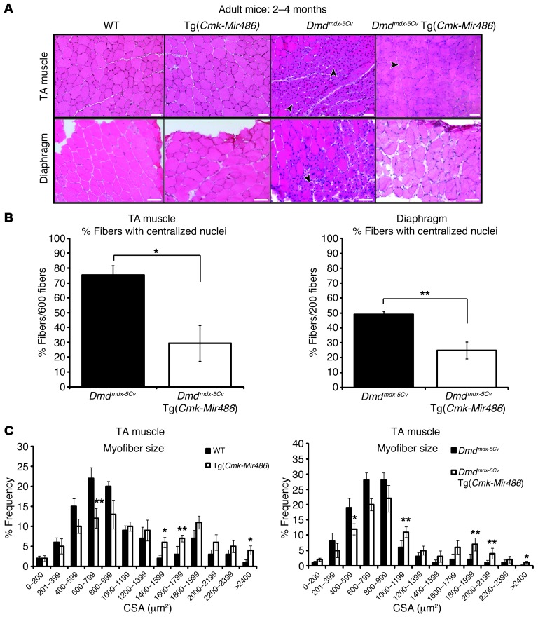
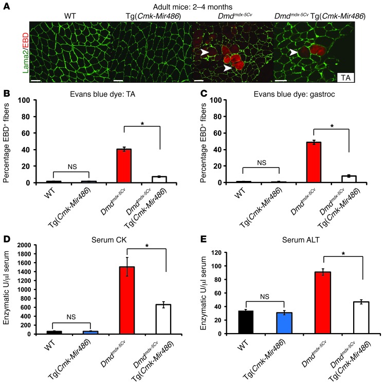
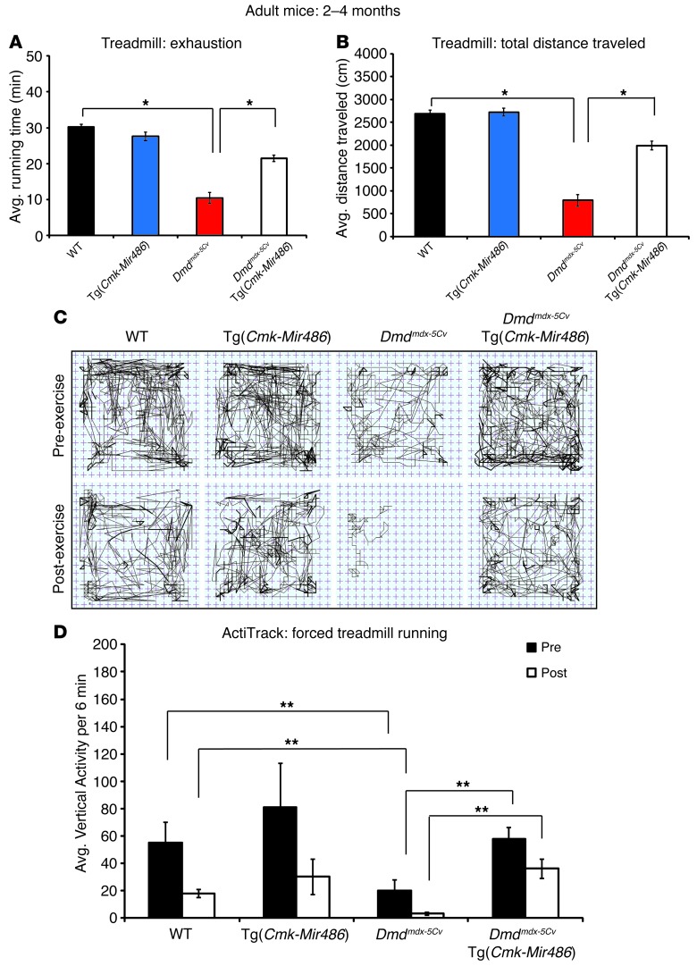
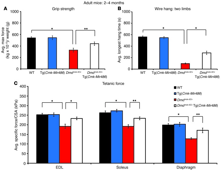
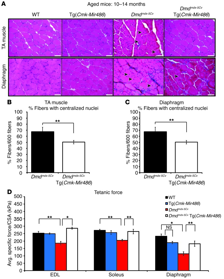
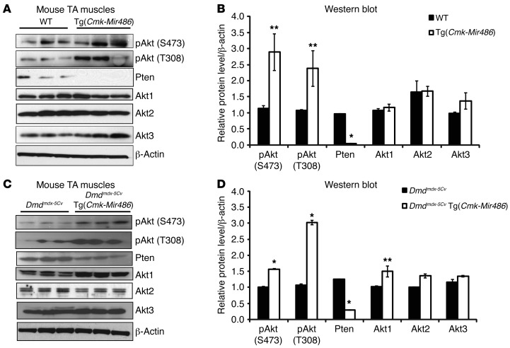
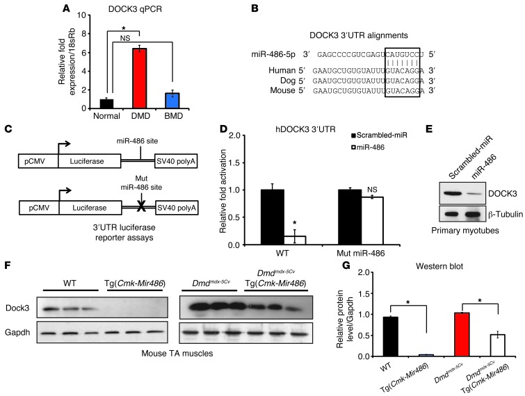
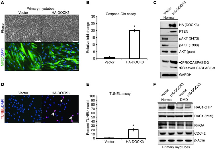
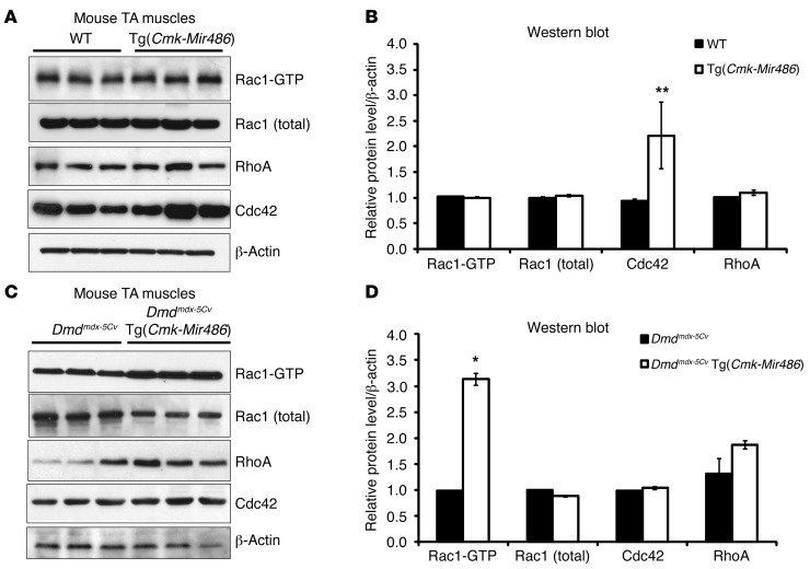
Duchenne muscular dystrophy (DMD) is caused by mutations in the gene encoding dystrophin, which results in dysfunctional signaling pathways within muscle. Previously, we identified microRNA-486 (miR-486) as a muscle-enriched microRNA that is markedly reduced in the muscles of dystrophin-deficient mice (Dmdmdx-5Cv mice) and in DMD patient muscles. Here, we determined that muscle-specific transgenic overexpression of miR-486 in muscle of Dmdmdx-5Cv mice results in reduced serum creatine kinase levels, improved sarcolemmal integrity, fewer centralized myonuclei, increased myofiber size, and improved muscle physiology and performance. Additionally, we identified dedicator of cytokinesis 3 (DOCK3) as a miR-486 target in skeletal muscle and determined that DOCK3 expression is induced in dystrophic muscles. DOCK3 overexpression in human myotubes modulated PTEN/AKT signaling, which regulates muscle hypertrophy and growth, and induced apoptosis. Furthermore, several components of the PTEN/AKT pathway were markedly modulated by miR-486 in dystrophin-deficient muscle. Skeletal muscle-specific miR-486 overexpression in Dmdmdx-5Cv animals decreased levels of DOCK3, reduced PTEN expression, and subsequently increased levels of phosphorylated AKT, which resulted in an overall beneficial effect. Together, these studies demonstrate that stable overexpression of miR-486 ameliorates the disease progression of dystrophin-deficient skeletal muscle.


Figures

Figure 1
Figure 1. Overexpression of miR-486 in dystrophic Dmdmdx-5Cv mouse muscles reduces dystrophic disease histopathology.
(A) H&E staining of adult TA and diaphragm muscles from WT, Tg(Cmk-Mir486), Dmdmdx-5Cv, and Dmdmdx-5Cv Tg(Cmk-Mir486) mice. Arrowheads demarcate centralized nuclei, a classic finding in dystrophic disease and regenerating skeletal muscle. Scale bars: 50 μm. (B) Summary graphs of centralized nuclei counted from the TA and diaphragm muscles of adult Dmdmdx-5Cv and Dmdmdx-5Cv Tg(Cmk-Mir486) mice. (C) Fiber type cross-section area analysis of the TA muscles of WT, Tg(Cmk-Mir486), Dmdmdx-5Cv, and Dmdmdx-5Cv Tg(Cmk-Mir486) adult mice. Six-hundred fibers from 5 separate mice per cohort were used for analysis. *P < 0.005; **P < 0.05. The mean and SEM values for the 4 genotype cohorts are as follows: WT (773 ± 27), Tg(Cmk-Mir486) (907 ± 19), Dmdmdx-5Cv(691 ± 42), and Dmdmdx-5Cv Tg(Cmk-Mir486) (815 ± 36) measured in μm2.
Figure 2
Figure 2. Dmdmdx-5Cv Tg(Cmk-Mir486) mice have improved muscle membrane integrity and serum biochemistry.
(A) EBD (red fluorescence) immunohistochemistry of TA muscle biopsies taken from WT, Tg(Cmk-Mir486), Dmdmdx-5Cv, and Dmdmdx-5Cv Tg(Cmk-Mir486) mice. Sections were costained with Laminin α-2 antisera (Lama2; green) to demarcate the myofiber basal lamina. Arrowheads demarcate EBD-positive myofibers. Scale bars: 50 μm. (B and C) Graph showing the quantification of EBD-positive myofibers from the TA (B) and gastrocnemius (C) muscles in WT (black bars), Tg(Cmk-Mir486) (blue bars), Dmdmdx-5Cv (red bars), and Dmdmdx-5Cv Tg(Cmk-Mir486) (white bars) mice. Six-hundred fibers from 3 separate mice per cohort were quantified. (D and E) Serum CK and ALT enzymatic levels (measured as enzymatic units per μl) taken from the 4 genotype cohorts (n = 5 mice per genotype cohort used). *P < 0.05. Gastroc, gastrocnemius.
Figure 3
Figure 3. miR-486 overexpression in Dmdmdx-5Cv mice improves dystrophic muscle physiology.
(A) Graph of forced downhill treadmill running of WT (black bars), Tg(Cmk-Mir486) (blue bars), Dmdmdx-5Cv (red bars), and Dmdmdx-5Cv Tg(Cmk-Mir486) (white bars) mice measuring time of endurance (total time before the mice reached exhaustion) in minutes. (B) Graph of forced downhill treadmill running measuring total distance traveled in centimeters (cm) of the 4 genotype cohorts. (C) Representative ActiTrack activity plots of the 4 mouse genotype cohorts measured before (pre-exercise) and after (post-exercise) forced downhill treadmill running. Thin lines represent mouse distance traveled, while darker lines represent instances of rearing (vertical activity). (D) Summary graph plotting the total number of instances of rearing (vertical activity) of mice before and after forced treadmill running. *P < 0.005; **P < 0.05.
Figure 4
Figure 4. miR-486–overexpressing dystrophic muscles have improved strength and tetanic force outputs.
(A) Cage-grip strength measurements of mice of the 4 genotype cohorts measured as kg × 10–5 and normalized to mouse weight (grams). (B) Two-limb (forelimbs) wire hang-time measurements (seconds) of each of the 4 genotype cohorts. (C) Tetanic muscle-force measurements, normalized to CSA, of isolated EDL, soleus, and diaphragm muscles. Eight (WT and Tg[Cmk-Mir486]) and 12 (Dmdmdx-5Cv and Dmdmdx-5Cv Tg[Cmk-Mir486]) adult (2 to 4 months old) male mice were used for each genotype cohort. *P < 0.005; **P < 0.05.
Figure 5
Figure 5. Aged dystrophic mice show similar improved pathology resulting from miR-486 transgenic overexpression.
(A) Representative H&E staining of TA and diaphragm muscles taken from aged (10 to 14 months old) WT, Tg(Cmk-Mir486), Dmdmdx-5Cv, and Dmdmdx-5Cv Tg(Cmk-Mir486) mice. Arrowheads represent centralized myonuclei. Scale bars: 50 μm. (B) Graphs of centralized myonuclei counts of the TA and (C) diaphragm muscles of aged Dmdmdx-5Cv and Dmdmdx-5Cv Tg(Cmk-Mir486) mice. Three mice (n = 6 TA and n = 3 diaphragm muscles) were used, with 600 TA and 200 diaphragm individual myofibers being counted. (D) Tetanic muscle-force measurements, normalized to CSA, of isolated EDL, soleus, and diaphragm muscles. Five (WT and Tg[Cmk-Mir486]) to 8 (Dmdmdx-5Cv and Dmdmdx-5Cv Tg[Cmk-Mir486]) aged (10 to 14 months old) male mice were used for each genotype cohort. *P < 0.005; **P < 0.05.
Figure 6
Figure 6. PTEN/AKT signaling is dysregulated in miR-486–overexpressing mouse muscle.
(A) Western blot images of whole-tissue lysates from adult WT and Tg(Cmk-Mir486) mouse TA muscles. Three mice were used per genotype cohort. (B) Western blot densitometry values of phospho-AKT (S473), phospho-AKT (T308), AKT1/2/3, and PTEN protein images normalized to both the first lane and the β-actin–loading control. (C) Western blot images of whole-tissue lysates from adult Dmdmdx-5Cv and Dmdmdx-5Cv Tg(Cmk-Mir486). Three mice were used per genotype cohort. (D) Western blot densitometry values of phospho-AKT (S473), phospho-AKT (T308), AKT1/2/3, and PTEN protein images normalized to both the first lane and the β-actin–loading control. *P < 0.005; **P < 0.05.
Figure 7
Figure 7. DOCK3 is a direct target of miR-486 in skeletal muscle.
(A) Real-time qPCR of human DOCK3 expression levels in normal, DMD, and BMD muscle biopsies. Expression levels are normalized to the 18sRb loading control. (B) Evolutionary conservation of the miR-486 seed site in mammalian DOCK3 3′ UTRs. Human, dog, and mouse are shown aligned with the seed region of miR-486-5p (boxed inset). (C) Schematic of 3′ UTR of the miR-486 target fused to a luciferase reporter construct. The miR-486 seed site is mutated in the mutant constructs. (D) Relative luciferase fold expression of the human DOCK3 3′ UTR fused to luciferase and transfected into HEK293T cells with either miR-486 or scrambled miR control plasmids. (E) Western blot analysis of human DOCK3 protein expression in primary human myotubes overexpressing either miR-486 or scrambled miR control lentivirus. β-Tubulin is shown as a loading control. (F) Western blot of DOCK3 protein in WT, Tg(Cmk-Mir486), Dmdmdx-5Cv, and Dmdmdx-5Cv Tg(Cmk-Mir486) TA muscle lysates. GAPDH is shown as a loading control. (G) Densitometry graph of DOCK3 protein expression normalized to either WT or Dmdmdx-5Cv levels,and the GAPDH loading control. *P < 0.005.
Figure 8
Figure 8. DOCK3 overexpression induces apoptosis and affects PTEN/AKT and RAC1/RHOA signaling in primary human myotubes.
(A) Phase microscopy images of primary human myotubes expressing either a control vector (pCI-Neo) or HA-DOCK3. Stable-transfected (vector or HA-DOCK3) primary myoblasts were differentiated into myotubes by adding differentiation medium for 72 hours prior to imaging. Note the large phase-bright cell aggregates in the DOCK3-overexpressing primary myotubes as compared with vector controls. Scale bars: 100 μm. (B) Activated caspase-3 is induced in primary human myotubes that overexpress DOCK3. Three replicates in separate wells were used for both DOCK3 and vector controls. (C) Western blot images showing DOCK3 overexpression affects PTEN/AKT signaling levels. Western blot images of HA (DOCK3), PTEN, pAKT (S473 and T308), AKT (pan/recognizes all 3 AKT isoforms), PROCASPASE3 (top arrowhead), cleaved CASPASE-3 (bottom arrowhead), and a GAPDH loading control taken from 30 μg of protein whole-cell lysates overexpressing either the control empty vector or HA-DOCK3 in primary human myotubes. (D) TUNEL assay on DOCK3-overexpressing and control vector–overexpressing primary human myotubes. The TUNEL-positive nuclei (red) are costained with DAPI (blue) in the vector (pCI-HA) control– and HA-DOCK3–overexpressing samples. (E) The percentage of TUNEL-positive myonuclei in the vector control– or HA-DOCK3–overexpressing normal primary myotubes is shown on the graph. Two-hundred myonuclei were counted from at least 10 different fields for each condition. Three separate 2-well chamber slides were used in this experiment (n = 6 wells per condition). *P < 0.005. (F) DOCK3 overexpression fails to induce active RAC1 (RAC1-GTP) in normal and DMD primary myotubes overexpressing either HA-DOCK3 or a control vector. Western blot images of other RAC1 downstream signaling factors (CDC42 and RHOA), DOCK3, and a β-actin–loading control are also shown.
Figure 9
Figure 9. RAC1 activation is induced in miR-486–overexpressing muscles.
(AD) The overexpression of miR-486 in dystrophic Dmdmdx-5Cv muscles results in increased levels of active Rac1 (Rac1-GTP). (A) Western blot images of whole-tissue lysates from adult (2 to 4 months old) WT and Tg(Cmk-Mir486) mouse TA muscles. Three mice were used per genotype cohort. (B) Densitometry graph of the Western blot images of whole-tissue lysates from adult WT and Tg(Cmk-Mir486) mouse TA muscles. (C) Western blot images of whole-tissue lysates from adult (2 to 4 months old) Dmdmdx-5Cv, and Dmdmdx-5Cv Tg(Cmk-Mir486) mouse TA muscles. (D) Densitometry graph of the Western blot images of whole-tissue lysates from adult WT and Tg(Cmk-Mir486) mouse TA muscles. Western blot protein levels were normalized to the total levels of the β-actin–loading control. *P < 0.005; **P < 0.05.

References

    1. Emery AE. The muscular dystrophies. Lancet. 2002;359(9307):687–695. doi: 10.1016/S0140-6736(02)07815-7. - DOI - PubMed
    1. Goemans NM, et al. Systemic administration of PRO051 in Duchenne’s muscular dystrophy. N Engl J Med. 2011;364(16):1513–1522. doi: 10.1056/NEJMoa1011367. - DOI - PubMed
    1. Cirak S, et al. Exon skipping and dystrophin restoration in patients with Duchenne muscular dystrophy after systemic phosphorodiamidate morpholino oligomer treatment: an open-label, phase 2, dose-escalation study. Lancet. 2011;378(9791):595–605. doi: 10.1016/S0140-6736(11)60756-3. - DOI - PMC - PubMed
    1. Liew WK, Kang PB. Recent developments in the treatment of Duchenne muscular dystrophy and spinal muscular atrophy. Ther Adv Neurol Disord. 2013;6(3):147–160. doi: 10.1177/1756285612472386. - DOI - PMC - PubMed
    1. Mendell J, et al. Eteplirsen for the treatment of duchenne muscular dystrophy. Ann Neurol. 2013;74(5):637–647. doi: 10.1002/ana.23982. - DOI - PubMed

Publication types

MeSH terms