Review
. 2014 May;29(3):203-15.
doi: 10.1152/physiol.00055.2013.

Alcohol abuse: critical pathophysiological processes and contribution to disease burden

Affiliations
Review

Alcohol abuse: critical pathophysiological processes and contribution to disease burden

Patricia E Molina et al. Physiology (Bethesda). 2014 May.

Abstract

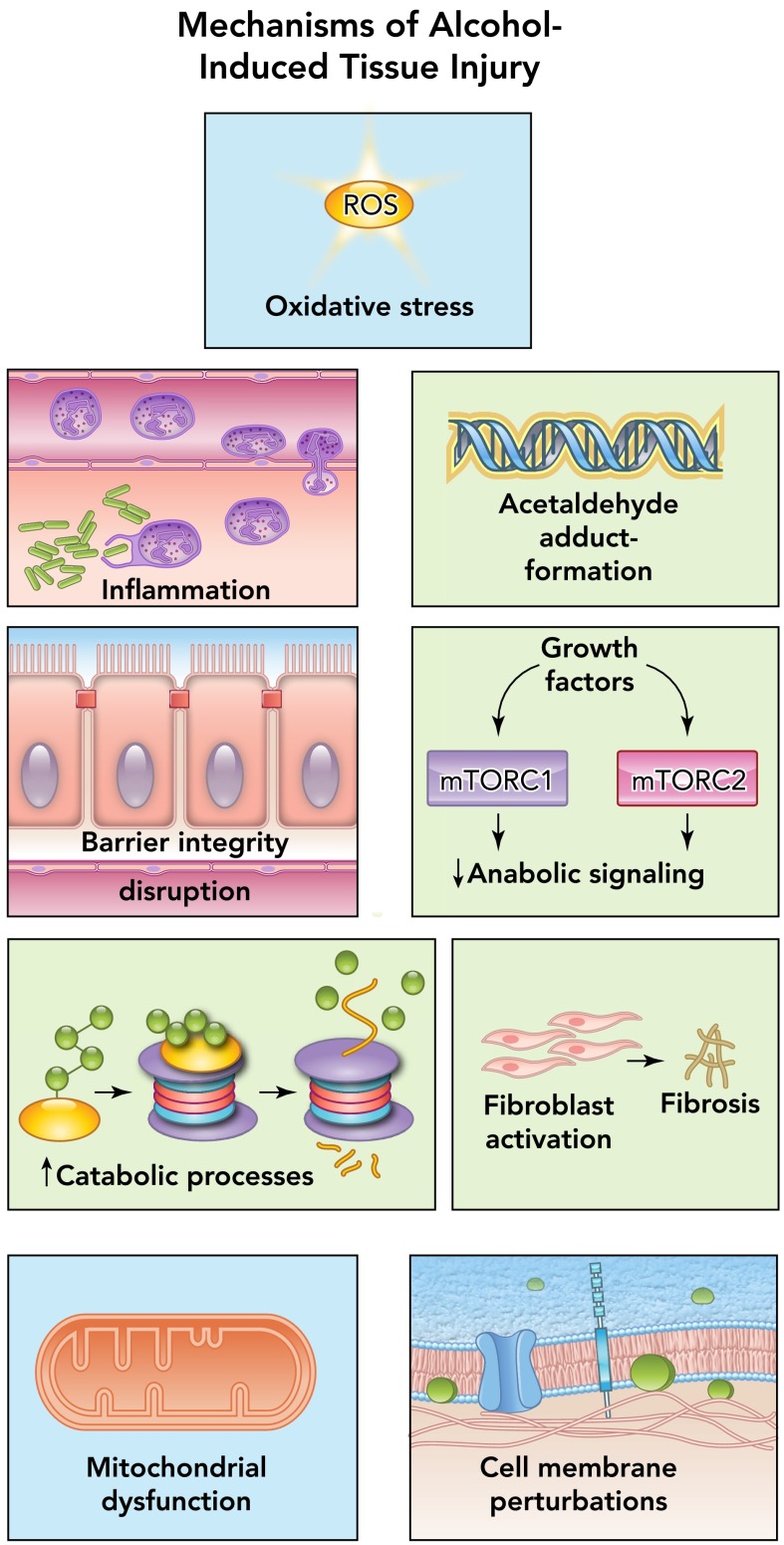
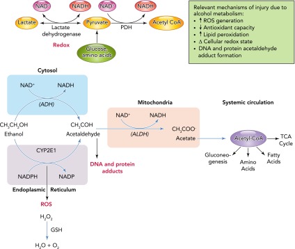
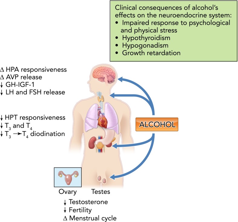
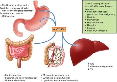
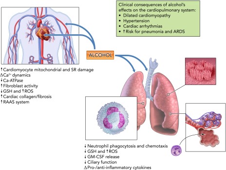
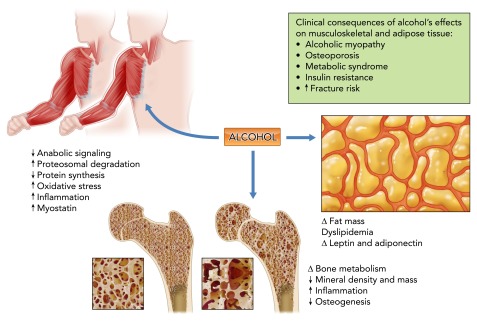
Alcohol abuse; the most common and costly form of drug abuse, is a major contributing factor to many disease categories. The alcohol-attributable disease burden is closely related to the average volume of alcohol consumption, with dose-dependent relationships between amount and duration of alcohol consumption and the incidence of diabetes mellitus, hypertension, cardiovascular disease, stroke, and pneumonia. The frequent occurrence of alcohol use disorders in the adult population and the significant and widespread detrimental organ system effects highlight the importance of recognizing and further investigating the pathophysiological mechanisms underlying alcohol-induced tissue and organ injury.


Conflict of interest statement

No conflicts of interest, financial or otherwise, are declared by the author(s).

Figures

FIGURE 1.
FIGURE 1.
Principal mechanisms of alcohol-induced pathophysiology Alcohol abuse disrupts multiple cellular mechanisms, leading to altered organ function and disease. Among the most important pathophysiological mechanisms identified as causative factors in tissue and organ injury resulting from alcohol abuse include oxidative stress, inflammation, acetaldehyde generation and adduct formation, decreased barrier function, impaired anabolic signaling, upregulation of catabolic processes, fibroblast activation, mitochondrial injury, and cell membrane perturbations.
FIGURE 2.
FIGURE 2.
Alcohol metabolism and its contribution to tissue injury The average rate at which alcohol is eliminated from the body is ∼7 g/h, which translates to ∼1 drink/h. Alcohol undergoes first pass gastric metabolism by the enzyme alcohol dehydrogenase (ADH). However, most alcohol oxidation occurs in the liver. Alcohol is metabolized to acetaldehyde primarily by alcohol dehydrogenase (ALD) and the cytochrome P450 2E1 (CYP2E1). This later pathway is particularly relevant following chronic alcohol abuse. Acetaldehyde is converted to acetate in the mitochondria by the enzyme acetaldehyde dehydrogenase (ALDH) type 2. Most of the acetate produced enters the systemic circulation and is activated to acetyl coenzyme A (CoA), a key intermediate metabolite in peripheral tissues. Acetaldehyde can form adducts that can produce injury through activation of immune responses. During the oxidative process, both ADH and ALDH reactions reduce NAD+ to NADH, shifting the cellular redox ratio, thereby affecting several NAD+ requiring enzymes like lactate and pyruvate dehydrogenase and affecting pathways including glycolysis, citric acid cycle, fatty acid oxidation, and gluconeogenesis. In addition, the cytochrome P450 enzymes, particularly CYP2E1, contribute to the oxidation of alcohol to acetaldehyde, particularly at increasing alcohol concentrations as well as following their induction by chronic alcohol abuse. Because CYP2E1 is involved in oxidation of several drugs to their reactive intermediates (e.g., nitrosamines, acetaminophen, and halothane), their toxicity is enhanced in alcoholics. This pathway of alcohol oxidation results in the production of large amounts of reactive oxygen species (ROS) and is thought to be an important mechanism contributing to alcoholic liver injury. ROS are eliminated by antioxidants like glutathione (GSH) under normal conditions. Alcohol depletes cellular GSH stores, thereby further exacerbating ROS-mediated injury. ROS can interact with lipids, producing lipid peroxidation, leading to formation of reactive molecules such as malondialdehyde (MDA) and 4-hydroxy-2-nonenal (HNE), which can in turn form protein adducts. A minor fraction of alcohol metabolism occurs in peroxisomes through catalase-dependent oxidation. Alcohol can also react with glucuronic acid to form ethyl-glucuronide, a soluble, non-volatile conjugate that is readily excreted and detected in body fluids, tissue, sweat, and hair for an extended time following alcohol consumption.
FIGURE 3.
FIGURE 3.
Alcohol and the neuroendocrine system Alcohol disrupts responsiveness of the hypothalamo-pituitary-adrenal (HPA) axis to psychological and physical stressors, and this has been implicated in the pathophysiology of pseudo-Cushing's syndrome, addiction, dependence, and relapse of recovering alcoholics. Alcohol produces dose-, frequency-, and duration-specific effects on arginine vasopressin (AVP), leading to alterations in water balance and mean arterial blood pressure homeostasis. Alcohol decreases the responsiveness of the hypothalamo-pituitary-thyroid (HPT) axis to central stimulation, decreases circulating levels of triiodothyronine (T3) and thyroxine (T4), and deiodination of T4 to T3. Chronic alcohol consumption disrupts the hypothalamo-pituitary-gonadal (HPG) axis and results in decreased testosterone levels, abnormal menstrual cycles, and infertility. GH, growth hormone; LH, luteinizing hormone; FSH, follicle stimulating hormone. The potential clinical consequences of alcohol abuse and its impact on the endocrine system are shown in the box.
FIGURE 4.
FIGURE 4.
Alcohol and the gut-liver axis Alcohol abuse produces marked alterations in the gastrointestinal tract. Esophageal and gastric dysmotility facilitate acid regurgitation and contribute to postemetic lacerations of the distal esophagus induced by vomiting (Mallory-Weiss Syndrome). Liver fibrosis and the resulting intrahepatic pressure increase leads to development of esophageal varicosities. Alcohol promotes chronic gastritis followed by decreased mucosal thickness and atrophy during the chronic phase. Chronic alcohol abuse impairs intestinal essential amino acid and vitamin absorption. In the liver, alcohol metabolism increases the production of ROS and lowers antioxidant levels, which contributes to liver injury. ROS generation leads to lipid peroxidation, alterations in plasma and intracellular membranes, and release of proinflammatory and profibrotic mediators. Alcohol and its metabolites disrupt intestinal barrier function by affecting the integrity of tight junctions, promoting the dissociation and redistributing proteins like ZO-1, claudin, and occludin. Increased paracellular permeability leads to increased bacterial toxin translocation from the gut lumen and disseminated to the systemic circulation via the portal vein and the lymphatic route. This later route of dissemination may be significant, since alcohol intoxication has been shown to promote lymphatic pumping. GSH, reduced glutathione; ROS, reactive oxygen species; HCV, hepatitis C virus; LES, lower esophageal sphincter. The potential clinical consequences of alcohol abuse and its impact on the endocrine system are shown in the box.
FIGURE 5.
FIGURE 5.
Alcohol and the cardiopulmonary system Chronic alcohol abuse increases risk for cardiovascular and pulmonary disease. Alcohol-induced myocardial dysfunction results from oxidative stress, cardiomyocyte mitochondrial and sarcoplasmic reticulum damage, altered calcium dynamics, and cardiac fibrosis. Alcohol abuse induces myocardial oxidative stress and depletes mitochondrial GSH, decreasing antioxidant capacity and enhancing myocyte susceptibility to oxidant injury and apoptosis, and in addition disrupts cardiomyocyte contraction by damaging contractile proteins and interfering with calcium signaling and homeostasis through upregulation of L-type calcium channel expression and function. Alcohol-mediated hypertension results from potentiation of the RAAS reflected in elevated circulating angiotensin II levels, cardiac angiotensin converting enzyme, and angiotensin type 1 (AT1) receptor expression. Chronic alcohol abuse produces marked alterations in pulmonary function resulting from decreased GSH, increased ROS production, and marked alterations in lung host defense mechanisms. GM-CSF, granulocyte-macrophage colony-stimulating factor; RAAS, renin-angiotensin-aldosterone system; ARDS, acute respiratory distress syndrome. The most relevant clinical manifestations of alcohol-induced alterations in cardiopulmonary function are shown in the box.
FIGURE 6.
FIGURE 6.
Alcohol and the musculoskeletal and adipose tissues Chronic alcohol abuse disrupts multiple factors involved the balance between anabolic and catabolic mechanisms in bone and muscle. The underlying mechanisms include nutritional deficiencies, decreased growth factor availability and responsiveness, increased ubiquitin proteasome pathway activation, upregulation of negative regulators of skeletal muscle growth, and disruption of bone remodeling. Chronic alcohol abuse produces marked alterations in adipocyte function, resulting in fat mass redistribution, dyslipidemia, and altered pattern of adipokine release. The potential clinical implications of alcohol's effects on skeletal muscle, bone, and adipose tissue are summarized in the box.

References

    1. Adachi Y, Moore LE, Bradford BU, Gao W, Thurman RG. Antibiotics prevent liver injury in rats following long-term exposure to ethanol. Gastroenterology 108: 218–224, 1995 - PubMed
    1. Addolorato G, Capristo E, Marini M, Santini P, Scognamiglio U, Attilia ML, Messineo D, Sasso GF, Gasbarrini G, Ceccanti M. Body composition changes induced by chronic ethanol abuse: evaluation by dual energy X-ray absorptiometry. Am J Gastroenterol 95: 2323–2327, 2000 - PubMed
    1. Adinoff B, Junghanns K, Kiefer F, Krishnan-Sarin S. Suppression of the HPA axis stress-response: implications for relapse. Alcohol Clin Exp Res 29: 1351–1355, 2005 - PMC - PubMed
    1. Aguilar D, Skali H, Moyé LA, Lewis EF, Gaziano JM, Rutherford JD, Hartley LH, Randall OS, Geltman EM, Lamas GA, Rouleau JL, Pfeffer MA, Solomon SD. Alcohol consumption and prognosis in patients with left ventricular systolic dysfunction after a myocardial infarction. J Am Coll Cardiol 43: 2015–2021, 2004 - PubMed
    1. Albano E, Vidali M. Immune mechanisms in alcoholic liver disease. Genes Nutr 5: 141–147, 2010 - PMC - PubMed

Publication types

LinkOut - more resources