. 2014 Jun 15;116(12):1582-92.
doi: 10.1152/japplphysiol.01362.2013. Epub 2014 May 1.

Age-related structural alterations in human skeletal muscle fibers and mitochondria are sex specific: relationship to single-fiber function

Affiliations

Age-related structural alterations in human skeletal muscle fibers and mitochondria are sex specific: relationship to single-fiber function

Damien M Callahan et al. J Appl Physiol (1985). .

Abstract

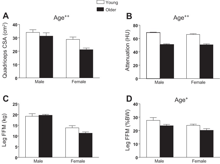
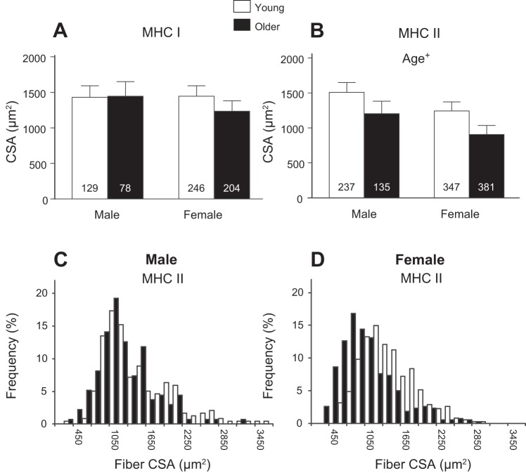
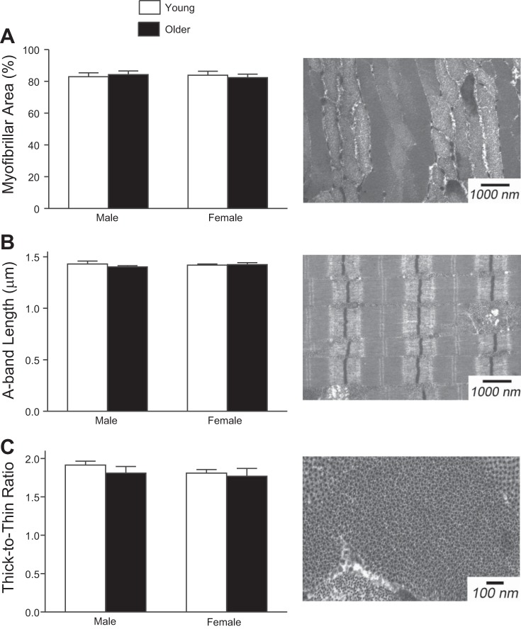
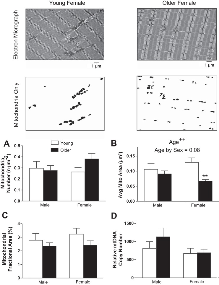
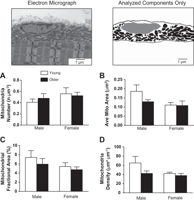
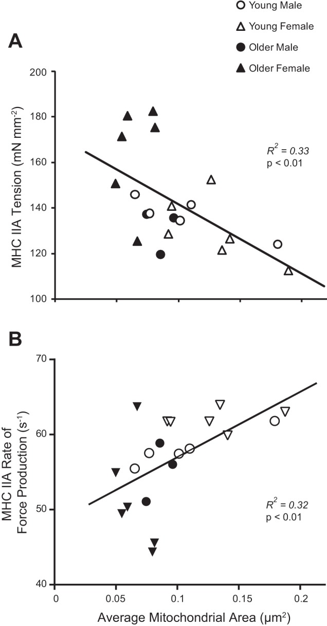
Age-related loss of skeletal muscle mass and function is implicated in the development of disease and physical disability. However, little is known about how age affects skeletal muscle structure at the cellular and ultrastructural levels or how such alterations impact function. Thus we examined skeletal muscle structure at the tissue, cellular, and myofibrillar levels in young (21-35 yr) and older (65-75 yr) male and female volunteers, matched for habitual physical activity level. Older adults had smaller whole muscle tissue cross-sectional areas (CSAs) and mass. At the cellular level, older adults had reduced CSAs in myosin heavy chain II (MHC II) fibers, with no differences in MHC I fibers. In MHC II fibers, older men tended to have fewer fibers with large CSAs, while older women showed reduced fiber size across the CSA range. Older adults showed a decrease in intermyofibrillar mitochondrial size; however, the age effect was driven primarily by women (i.e., age by sex interaction effect). Mitochondrial size was inversely and directly related to isometric tension and myosin-actin cross-bridge kinetics, respectively. Notably, there were no intermyofibrillar or subsarcolemmal mitochondrial fractional content or myofilament ultrastructural differences in the activity-matched young and older adults. Collectively, our results indicate age-related reductions in whole muscle size do not vary by sex. However, age-related structural alterations at the cellular and subcellular levels are different between the sexes and may contribute to different functional phenotypes in ways that modulate sex-specific reductions in physical capacity with age.

Keywords: aging; mitochondria; muscle ultrastructure; sarcopenia.


Figures

Fig. 1.
Fig. 1.
Whole muscle morphology was characterized using computerized tomography (CT; A and B) and dual-energy X-ray absorptiometry (DEXA; C and D). A: quadriceps cross-sectional area (CSA). B: attenuation in Hounsfield units (HU). C and D: leg fat-free mass in kg (C) and %body weight (%BW; D). Values are means ± SE. Text indicates significant difference (+P ≤ 0.05 or ++P < 0.01) between young and older groups (Age).
Fig. 2.
Fig. 2.
Single-fiber CSA from myosin heavy chain (MHC) I (A) and MHC II (B) fiber types determined using immunohistochemical analysis. Text indicates significant difference (+P < 0.05) between young and older groups (Age). Nos. in bars in A and B indicate the no. of measured fibers. Values are means ± SE. The relative frequency of muscle fiber CSA in MHC II fibers is presented for men (C) and women (D).
Fig. 3.
Fig. 3.
Myofibrillar fractional area (A), A-band length (B), and thick-to-thin-filament ratio (C) were assessed using transmission electron microscopy (EM). These indexes of muscle ultrastructure were not different between young and older men and women. Example images from a young woman are presented to the right of the corresponding data.
Fig. 4.
Fig. 4.
Intermyofibrillar mitochondrial fractional area and morphology were assessed using transmission EM. Representative EM images (top) are shown with contrasted versions of the same image below, which were used to identify mitochondrial number (n·μm−2; A), average mitochondrial CSA (μm2; B), and mitochondrial density or fractional area (%field of view; C). D: relative mitochondrial DNA (mtDNA) copy number. Values are means ± SE. Text indicates significant difference (++P < 0.01) between young and older groups (Age) and a trend between age and sex (Age by Sex). **Significant pairwise differences (P < 0.01) between young and older women.
Fig. 5.
Fig. 5.
Subsarcolemmal mitochondrial fractional area and morphology were assessed using transmission EM, similar to the intermyofibrillar mitochondria (Fig. 4). Top: a representative EM image is shown with an image of its contrasted mitochondria, used to identify morphological characteristics. Subsarcolemmal mitochondria were those between myofibrils and the sarcolemma (indicated by dashed lines). The nucleus in the subsarcolemmal region is indicated in gray and was not included in the area analysis of the subsarcolemmal space. A: mitochondrial number. B: average mitochondrial area. C: mitochondrial fractional area. D: mitochondrial density. Values are means ± SE.
Fig. 6.
Fig. 6.
Mitochondrial morphology (average mitochondrial CSA) was significantly correlated with average single-fiber Ca2+ activated tension (A) and rate of force development (B) in MHC IIA fibers.

Similar articles

Cited by

References

    1. Ahtiainen M, Pollanen E, Ronkainen PH, Alen M, Puolakka J, Kaprio J, Sipila S, Kovanen V. Age and estrogen-based hormone therapy affect systemic and local IL-6 and IGF-1 pathways in women. Age (Dordr) 34: 1249–1260, 2012 - PMC - PubMed
    1. Bertoni-Freddari C, Fattoretti P, Caselli U, Giorgetti B, Albanelli S, Torelli F, Felzani G, Vecchiet J. A morphometric study on human muscle mitochondria in aging. J Am Aging Assoc 25: 101–105, 2002 - PMC - PubMed
    1. Brierly EJ, Johnson MA, Bowman A, Ford GA, Subhan F, Reed JW, James OF, Turnbull DM. Mitochondrial function in muscle from elderly athletes. Ann Neurol 41: 114–116, 1997 - PubMed
    1. Broskey NT, Greggio C, Boss A, Boutant M, Dwyer A, Schlueter L, Hans D, Gremion G, Kreis R, Boesch C, Canto C, Amati F. Skeletal muscle mitochondria in the elderly: effects of physical fitness and exercise training. J Clin Endocrinol Metab 99: 1852–1861, 2014 - PubMed
    1. Callahan DM, Kent-Braun JA. Effect of old age on human skeletal muscle force-velocity and fatigue properties. J Appl Physiol 111: 1345–1352, 2011 - PMC - PubMed

Publication types