. 2014 Jun 1;192(11):4967-76.
doi: 10.4049/jimmunol.1301670. Epub 2014 Apr 30.

Distinct assembly profiles of HLA-B molecules

Affiliations

Distinct assembly profiles of HLA-B molecules

Syed Monem Rizvi et al. J Immunol. .

Abstract

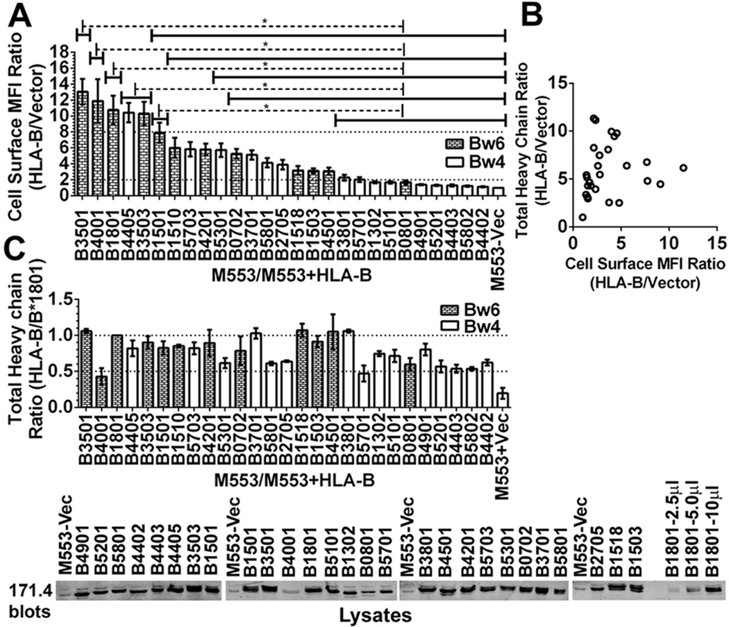
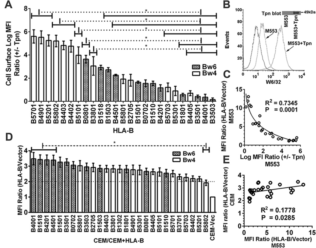
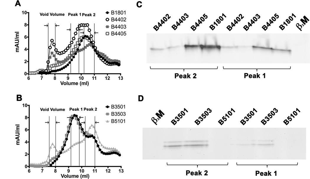
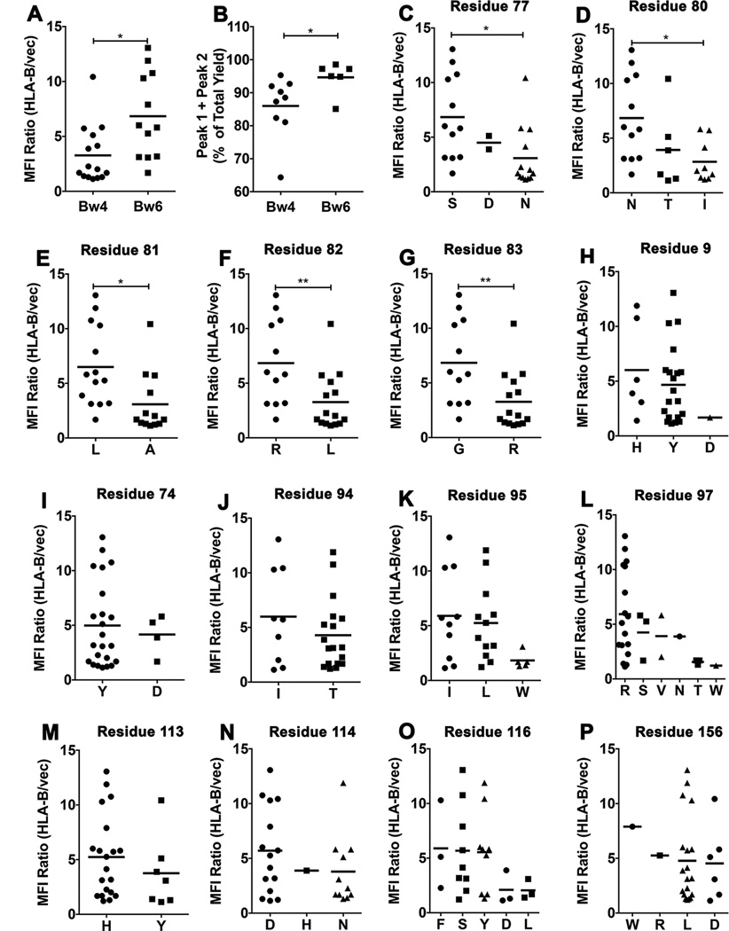
MHC class I polymorphisms are known to influence outcomes in a number of infectious diseases, cancers, and inflammatory diseases. Human MHC class I H chains are encoded by the HLA-A, HLA-B, and HLA-C genes. These genes are highly polymorphic, with the HLA-B locus being the most variable. Each HLA class I protein binds to a distinct set of peptide Ags, which are presented to CD8(+) T cells. HLA-disease associations have been shown in some cases to link to the peptide-binding characteristics of individual HLA class I molecules. In this study, we show that polymorphisms at the HLA-B locus profoundly influence the assembly characteristics of HLA-B molecules and the stabilities of their peptide-deficient forms. In particular, dependence on the assembly factor tapasin is highly variable, with frequent occurrence of strongly tapasin-dependent or independent allotypes. Several polymorphic HLA-B residues located near the C-terminal end of the peptide are key determinants of tapasin-independent assembly. In vitro refolded forms of tapasin-independent allotypes assemble more readily with peptides compared to tapasin-dependent allotypes that belong to the same supertype, and, during refolding, reduced aggregation of tapasin-independent allotypes is observed. Paradoxically, in HIV-infected individuals, greater tapasin-independent HLA-B assembly confers more rapid progression to death, consistent with previous findings that some HLA-B allotypes shown to be tapasin independent are associated with rapid progression to multiple AIDS outcomes. Together, these findings demonstrate significant variations in the assembly of HLA-B molecules and indicate influences of HLA-B-folding patterns upon infectious disease outcomes.


Figures

Figure 1
Figure 1. Variable expression of HLA-B molecules in tapasin-deficient cells
A) The y-axes show cell surface expression ratios (mean fluorescence intensity (MFI ratios)) of MHC class I staining in M553 cells (a tapasin-deficient melanoma cell line) infected with retroviral constructs encoding the indicated HLA-B allotypes relative to M553 cells that were infected with a virus lacking MHC class I (vector). MHC class I surface expression was analyzed by flow cytometry using the W6/32 antibody. Statistical analyses were done using a one-way ANOVA test, followed by a Tukey’s multiple comparisons procedure for all pairwise differences of means. Significant differences are indicated (with an asterisk) on the graph (P< 0. 05). Data represents averaged MFI ratios derived from 10–15 independent flow cytometric analyses from five independent infections (infections 1–5) of M553 cells. B) In infection 5, a Pearson analysis was used to examine correlation between cell surface MFI ratios assessed by flow cytometry and total cellular expression ratios assessed by immunoblotting of cell lysates. A significant correlation was not observed. C) Top panel: Cell lysates from infection 5 of M553 cells were tested by quantitative fluorescence-based immunoblotting with the heavy chain-specific 171. 4 antibody. The bar graphs show total cellular expression ratios (relative to cells expressing HLA-B*1801) of the indicated HLA-B molecules. Averaged values from three independent sets of immunoblotting analyses of infection 5 are shown. Lower panel: Representative quantitative immunoblots with the 171. 4 antibody of indicated cell lysates from infection 5. 10 µl of total cell lysate was loaded in each lane, unless otherwise indicated. The last three lanes show HLA-B*1801 heavy chain expression in M553-HLA-B*1801 lysates (incremental loads of 2. 5–10 µl).
Figure 2
Figure 2. Reduced variability of HLA-B expression in tapasin-sufficient cells
A) Cells from one of the infections described in Fig. 1A were subsequently infected with a tapasin-encoding virus. Following subtractions of the vector alone backgrounds the MFI ratios in the presence and absence of tapasin (+tapasin/−tapasin MFI ratios) were calculated for each HLA-B-expressing cell line. B) Representative histograms of M553 cell infections with a tapasin-encoding or control retrovirus. Flow cytometric analyses with the W6/32 antibody of cell surface expression of the endogenous MHC class I of M553 cells that were infected with retroviruses encoding or lacking tapasin. C) A Pearson analysis was used to examine correlation between averaged cell surface MFI ratios (HLA-B/vector) in M553 (Fig. 1A) vs. Log MFI (+Tapasin/−Tapasin) ratios (Fig. 2A). D) CEM cells (a CD4 T cell line) were infected with retroviruses encoding indicated HLA-B allotypes or a control virus lacking HLA-B (Vector). MHC class I cell surface expression was analyzed by flow cytometry using the W6/32 antibody, and HLA-B/vector MFI ratios were determined. E) A Pearson analysis was used to examine correlation between averaged MFI ratios (HLA-B/vector) in M553 and CEM cells. Data represent averaged (A and D) MFI values derived from 2–4 (A) or 10–12 (D) independent flow cytometric analyses from one (A) or three (D) independent infections. Statistical analyses (A and D) were done using a one-way ANOVA test, followed by a Tukey’s multiple comparisons procedure for all pairwise differences of means. Pearson R square (R2) and significantly different values (P) are indicated on the graph. P< 0. 05 is considered significant.
Figure 3
Figure 3. Higher assembly competence of soluble refolded tapasin-independent HLA-B allotypes
A soluble form of the indicated HLA class I heavy chains (2 µM) was refolded with β2m (4 µM) overnight at 4 °C in the presence of TEV. Following refolding, the soluble fractions were analyzed by gel filtration chromatography using a Superdex 75 10/300 GL column. Representative gel filtration analyses of A) B1801, B4402, B4403 and B4405 (B44 supertype) and B) B3501, B3503 and B5101 (B7 supertype) are shown. The vertical lines show peak position ranges of the gel filtration column void volume (left), Peak 1 (middle) and Peak 2 (right). C and D) Peak 1 and Peak 2 fractions from A and B were assessed for their peptide binding competence. Peak 1 and Peak 2 fractions were normalized for protein concentration (to 200 nM (Peak 1) or 400 nM (peak 2) for B44 supertypes, and 20 nM (peaks 1 and 2) for B7 supertypes) and incubated with a 10-fold excess of refolded β2m and FITC labeled peptide (peptide EEFGKFITCAFSF for B44 supertypes and peptide IPLKFITCEEAEL for B7 supertypes). Peptide bound heterodimers were separated by native gel electrophoresis and visualized by fluorescence scanning. Both Peak 1 and Peak 2 fractions are competent for peptide binding. Representative data of four independent measurements are shown.
Figure 4
Figure 4. Variable levels of aggregation of soluble empty HLA-B molecules following refolding
Soluble forms of the indicated MHC class I heavy chains were refolded with β2m A) overnight at 4 °C with TEV or B) at room temperature (22 ° C) for 1 hour without TEV. Following refolding, soluble proteins were analyzed by gel filtration chromatography. Percentages of fractions corresponding to the void volume and Peak1 + Peak 2 (peptide binding competent fractions) of different allotypes are shown (left Y axis) and total protein yields (right Y axis) are also shown. Data represent averaged values from three independent refolding reactions with two different inclusion body preparations for all allotypes except HLA-B*0801 and HLA-B*1302, which represent averaged values of two independent refolding reactions with a single inclusion body preparation. C and D) Percentages of Peak 1+ Peak 2 fractions, derived from data in A and B, were compared for the tapasin-independent and tapasin-dependent allotypes. Differences between two groups are significant when refolding is conducted under the more stringent conditions (1 hour at 22 °C). Statistical analyses were performed using an unpaired t-test. ** Indicates that the P value was < 0. 01.
Figure 5
Figure 5. Residue contributions to tapasin-independent assembly
A) Averaged MFI ratios derived in Fig. 1A were compared for HLA-Bw4 and HLA-Bw6 allotypes. Statistical analyses are based on an unpaired t-test. B) Peak 1+ Peak 2 yields, derived from the 4 ° C refolding data in Fig. 4A were compared for HLA-Bw4 and HLA-Bw6 allotypes. Statistical analyses are based on an unpaired t-test. More significant differences were observed for the 1 hour/room temperature refolding data. C–P) HLA-B allotypes were grouped based on the presence of dimorphic or polymorphic residues at the indicated heavy chain sequence positions. Averaged MFI ratios derived in Fig. 1A are compared for allotypes with a given amino acid residue at each of the indicated dimorphic or polymorphic sites. Statistical differences between groups were assessed based on unpaired t-tests for dimorphic residues. For polymorphic residues, a one-way ANOVA test was used, with a Tukey’s multiple comparisons procedure for all pairwise differences of means. Statistically significant differences are indicated by asterisks on the graphs (* indicates P< 0. 05; ** indicates P<0. 01).
Figure 6
Figure 6. Determinants of tapasin-independent assembly
A) Sequence alignments of HLA-B*4405 with tapasindependent HLA-Bw4 molecules and of HLA-B*0801 with tapasin-independent HLA-Bw6 molecules. B and C) Based on the sequence alignments, residues expected to contribute to tapasin-dependent and tapasin-independent HLA-B assembly are highlighted on the structures of HLA-B*4405 (pdb 1SYV) and HLA-B*0801 (pdb 1AGD).

Similar articles

Cited by

References

    1. Neefjes J, Jongsma ML, Paul P, Bakke O. Towards a systems understanding of MHC class I and MHC class II antigen presentation. Nat Rev Immunol. 2011;11:823–836. - PubMed
    1. Campbell KS, Purdy AK. Structure/function of human killer cell immunoglobulin-like receptors: lessons from polymorphisms, evolution, crystal structures and mutations. Immunology. 2011;132:315–325. - PMC - PubMed
    1. Carrington M, Walker BD. Immunogenetics of spontaneous control of HIV. Annu Rev Med. 2012;63:131–145. - PMC - PubMed
    1. Goulder PJ, Walker BD. HIV and HLA class I: an evolving relationship. Immunity. 2012;37:426–440. - PMC - PubMed
    1. Jamil KM, Khakoo SI. KIR/HLA interactions and pathogen immunity. J Biomed Biotechnol. 2011;2011:298348. - PMC - PubMed

Publication types

MeSH terms