. 2014 Aug;25(8):1662-8.
doi: 10.1681/ASN.2013040425. Epub 2014 May 1.

Administration of recombinant soluble urokinase receptor per se is not sufficient to induce podocyte alterations and proteinuria in mice

Affiliations

Administration of recombinant soluble urokinase receptor per se is not sufficient to induce podocyte alterations and proteinuria in mice

Dominique Cathelin et al. J Am Soc Nephrol. 2014 Aug.

Abstract

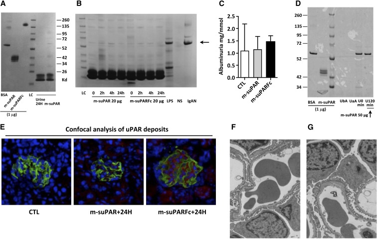
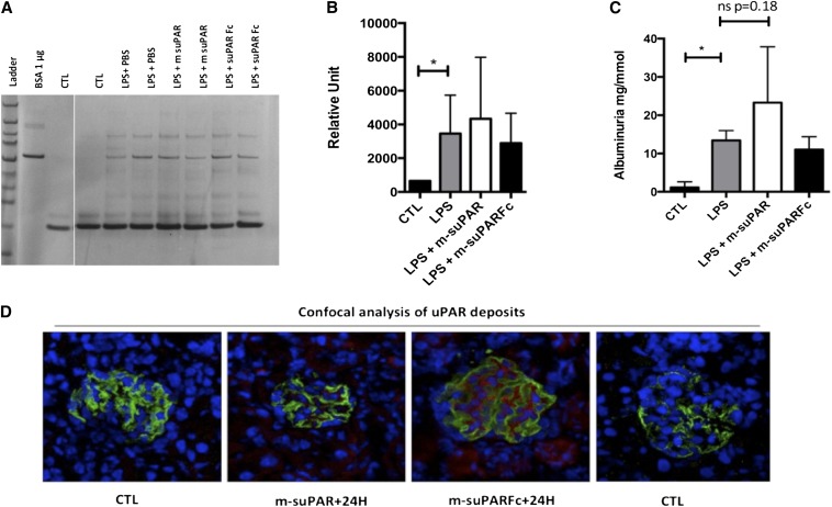
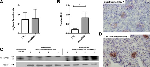
Circulating levels of soluble forms of urokinase-type plasminogen activator receptor (suPAR) are generally elevated in sera from children and adults with FSGS compared with levels in healthy persons or those with other types of kidney disease. In mice lacking the gene encoding uPAR, forced increases in suPAR concentration result in FSGS-like glomerular lesions and proteinuria. However, whether overexpression of suPAR, per se, contributes to the pathogenesis of FSGS in humans remains controversial. We conducted an independent set of animal experiments in which two different and well characterized forms of recombinant suPAR produced by eukaryotic cells were administered over the short or long term to wild-type (WT) mice. In accordance with the previous study, the delivered suPARs are deposited in the glomeruli. However, such deposition of either form of suPAR in the kidney did not result in increased glomerular proteinuria or altered podocyte architecture. Our findings suggest that glomerular deposits of suPAR caused by elevated plasma levels are not sufficient to engender albuminuria.

Keywords: glomerulosclerosis; nephrotic syndrome; pathophysiology of renal disease and progression; podocyte; proteinuria.


Figures

Figure 1.
Figure 1.
Intravenous injections of two different forms of eukaryotic recombinant mouse suPAR in wild-type (WT) mice (C57BL/6J or 129S2SvPasCrl, females 8–12 weeks old) do not cause foot process effacement or significant proteinuria despite the deposition of both recombinant proteins on glomerular structures. (A) Left: SDS-PAGE migration of purified recombinant mouse uPAR proteins (1 µg per lane) expressed by eukaryote cells: monomeric mouse uPAR produced in S2 cells (m-suPAR) and a commercially available uPAR-Fc chimera used in the study by Wei et al. The electrophoretic mobility of m-suPAR and m-suPARFc correspond to their expected molecular masses. Right: Injection of massive doses of these recombinant suPARs (100 µg) does not induce significant proteinuria in mice after 24 hours. LC, loading control. (B) Representative SDS-PAGE analyses of void urine normalized for creatinine content (1 mg per lane enables a strict comparison of the albuminuria development following injection of native [m-suPAR] or chimeric [m-suPARFc] soluble uPAR [20 µg equals the dose used in Wei et al.]). Urine from a normal subject (NS) without albuminuria serves as negative control, and urine specimens from a patient with IgA nephropathy (Berger's disease) and non–nephrotic-range proteinuria (15 mg/g creatinine) and from an LPS-treated mouse served as positive controls. The arrow indicates the electrophoretic mobility of albumin. (C) ELISA assessment of quantity of albuminuria 24 hours after uPAR administration (n=5–8 per group). CTL, control. (D) Invasive monitoring of bladder urine for development of proteinuria on a shorter time scale (2 hours) excludes the existence of transient albuminuria caused by circulating suPAR. Comparison between urine collected before suPAR administration (time zero, U0) and 120 minutes after injection (U120) are shown. Surgical stress and catheterization lead to the induction of spurious albuminuria, which is not related to the anesthesia procedure (urine before [UbA] and after [UaA] anesthesia). Image representative of four catheterized mice. On the left, 1 μg of either BSA or m-suPAR have been deposited as positive controls. (E) Confocal microscopy showing suPAR deposits on normal glomerular structures at 24 hours from mice depicted in B (suPAR in red and podocin in green). (F and G) Transmission electronic microscopy of mouse glomeruli after native suPAR (right) or chimeric suPAR (left) injection as depicted in A and B. No features of podocyte dedifferentiation are observed at 24 hours despite suPAR or chimeric suPAR deposits in E. All results are expressed as mean±SD (n=5–8 per group).
Figure 2.
Figure 2.
Intraperitoneal administration of two distinct forms of recombinant mouse suPAR 24 hours after the last LPS insult fails to exacerbate LPS-associated albuminuria. (A) Injection of 25 µg of either form of suPAR does not lead to further increases in albuminuria, as evaluated by density scanned SDS-PAGE gel (B) or ELISA (C) (n=8 per group). CTL, control. (D) Confocal microscopy demonstrates the increase in glomeruli expressed uPAR or sequestered soluble uPAR following LPS injection (suPAR/uPAR in red and podocin in green). On the right, CTL indicates staining without primary uPAR antibody, showing the absence of a nonspecific staining. All results are expressed as mean±SD (n=8 per group); *P<0.05 (one way ANOVA).
Figure 3.
Figure 3.
Long-term delivery of suPAR in WT mice. Seven-day sustained delivery of recombinant mouse suPAR (200 µg) by mini-osmotic pumps in normal WT mice (C57BL6/J). (A) Albuminuria (ELISA) evaluated after 7 days of mouse suPAR delivery; absence of any induction of albuminuria between NaCl-treated control (CTL) group versus the suPAR-delivered group (n=8–10 mice per group). (B and C) Western blot detection of uPAR in lysates of kidney cortex 7 days after mini-pump installation showing a massive accumulation of suPAR in the kidney (n=4 per group). The arrows indicate suPAR, and the double band aspect represents the endogenous glycosylation heterogeneity of mouse uPAR. (D) Immunohistochemical detection of suPAR deposited in the glomeruli of mouse kidneys 7 days after mini-pump installation. Deposits are found essentially on glomerular structures (magnification ×400). Overall, the subcutaneous delivery of 200 µg of recombinant suPAR over a week failed to induce any albuminuria in normal mice despite suPAR deposits/accumulation on glomerular structures. All results are expressed as mean±SD (n=8 per group); *P<0.05 (one-way ANOVA).

Comment in

References

    1. Hoyer JR, Vernier RL, Najarian JS, Raij L, Simmons RL, Michael AF: Recurrence of idiopathic nephrotic syndrome after renal transplantation. Lancet 2: 343–348, 1972 - PubMed
    1. Le Berre L, Godfrin Y, Perretto S, Smit H, Buzelin F, Kerjaschki D, Usal C, Cuturi C, Soulillou JP, Dantal J: The Buffalo/Mna rat, an animal model of FSGS recurrence after renal transplantation. Transplant Proc 33: 3338–3340, 2001 - PubMed
    1. Le Berre L, Godfrin Y, Günther E, Buzelin F, Perretto S, Smit H, Kerjaschki D, Usal C, Cuturi C, Soulillou JP, Dantal J: Extrarenal effects on the pathogenesis and relapse of idiopathic nephrotic syndrome in Buffalo/Mna rats. J Clin Invest 109: 491–498, 2002 - PMC - PubMed
    1. Wei C, Möller CC, Altintas MM, Li J, Schwarz K, Zacchigna S, Xie L, Henger A, Schmid H, Rastaldi MP, Cowan P, Kretzler M, Parrilla R, Bendayan M, Gupta V, Nikolic B, Kalluri R, Carmeliet P, Mundel P, Reiser J: Modification of kidney barrier function by the urokinase receptor. Nat Med 14: 55–63, 2008 - PubMed
    1. Wei C, El Hindi S, Li J, Fornoni A, Goes N, Sageshima J, Maiguel D, Karumanchi SA, Yap HK, Saleem M, Zhang Q, Nikolic B, Chaudhuri A, Daftarian P, Salido E, Torres A, Salifu M, Sarwal MM, Schaefer F, Morath C, Schwenger V, Zeier M, Gupta V, Roth D, Rastaldi MP, Burke G, Ruiz P, Reiser J: Circulating urokinase receptor as a cause of focal segmental glomerulosclerosis. Nat Med 17: 952–960, 2011 - PMC - PubMed

Publication types

MeSH terms

Substances