. 2014 Apr 30;34(18):6334-43.
doi: 10.1523/JNEUROSCI.4044-13.2014.

GPR179 is required for high sensitivity of the mGluR6 signaling cascade in depolarizing bipolar cells

Affiliations

GPR179 is required for high sensitivity of the mGluR6 signaling cascade in depolarizing bipolar cells

Thomas A Ray et al. J Neurosci. .

Abstract

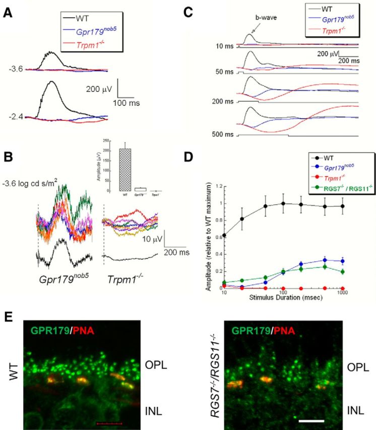
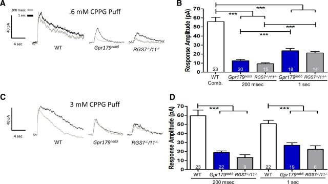
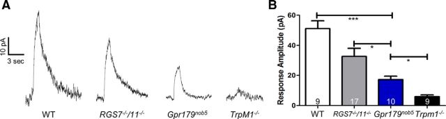
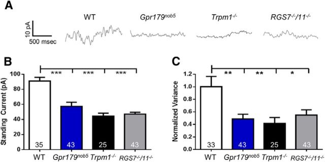
Parallel visual pathways are initiated at the first retinal synapse by signaling between the rod and cone photoreceptors and two general classes of bipolar cells. For normal function, ON or depolarizing bipolar cells (DBCs) require the G-protein-coupled receptor, mGluR6, an intact G-protein-coupled cascade and the transient receptor potential melastatin 1 (TRPM1) cation channel. In addition, another seven transmembrane protein, GPR179, is required for DBC function and recruits the regulators of G-protein signaling (RGS) proteins, RGS7 and RGS11, to the dendritic tips of the DBCs. Here we use the Gpr179(nob5) mouse, which lacks GPR179 and has a no b-wave electroretinogram (ERG) phenotype, to demonstrate that despite the absence of both GPR179 and RGS7/RGS11, a small dark-adapted ERG b-wave remains and can be enhanced with long duration flashes. Consistent with the ERG, the mGluR6-mediated gating of TRPM1 can be evoked pharmacologically in Gpr179(nob5) and RGS7(-/-)/RGS11(-/-) rod BCs if strong stimulation conditions are used. In contrast, direct gating of TRPM1 by capsaicin in RGS7(-/-)/RGS11(-/-) and WT rod BCs is similar, but severely compromised in Gpr179(nob5) rod BCs. Noise and standing current analyses indicate that the remaining channels in Gpr179(nob5) and RGS7(-/-)/RGS11(-/-) rod BCs have a very low open probability. We propose that GPR179 along with RGS7 and RGS11 controls the ability of the mGluR6 cascade to gate TRPM1. In addition to its role in localizing RGS7 and RGS11 to the dendritic tips, GPR179 via a direct interaction with the TRPM1 channel alters its ability to be gated directly by capsaicin.

Keywords: GPR179; TRPM1; mGluR6; night blindness; retina; rod bipolar.


Figures

Figure 1.
Figure 1.
mGluR6, TRPM1, and NYX expression is independent of GPR179 expression. Representative confocal images of cross sections of the OPL and inner nuclear layer (INL) of WT (A) and Gpr179nob5 (B) retinas reacted with antibodies to TRPM1 (green), mGluR6 (blue), and EYFP-Nyx (red). The merged images (bottom) show that expression patterns are similar in WT and in Gpr179nob5 retinas and that TRPM1, mGluR6, and nyctalopin expression colocalize at the dendritic tips of DBCs. Scale bars: 5 μm.
Figure 2.
Figure 2.
GPR179 expression at the dendritic tips of the DBCs is independent of mGluR6, TRPM1, and nyctalopin expression. Representative confocal images of cross sections of the OPL and inner nuclear layer (INL) of WT and a series of mutant mouse retinas reacted with an antibody to GPR179 (green) and the cone pedicle marker, PNA (red). A, In all retinas except Gpr179nob5, expression of GPR179 (green puncta) is found both at the rod BC dendritic tips and colocalized with PNA (yellow puncta in merged images). B–D, GPR179 expression is localized at the dendritic tips of DBCs even in the absence of expression of mGluR6 (B), nyctalopin (C), and TRPM1 (D). As shown here (E) and previously (Peachey et al., 2012b), GPR179 expression is absent from Gpr179nob5 mouse retina. Scale bars: 5 μm.
Figure 3.
Figure 3.
GPR179 and TRPM1 proteins interact. Western blots of total retinal lysates probed with antibodies to (A) GPR179 and (C) TRPM1 (top). Each blot was reprobed with antibodies to β-actin (bottom) to determine total protein and for use as an internal standard. Band intensities were analyzed and quantified with NIH ImageJ software and normalized to β-actin expression level in the same sample. The histograms (B, D) plot the mean expression (±SEM) from four experiments on independent retina samples. B, GPR179 expression was lower in Grm6−/− and Trpm1−/− retinas compared with WT. D, TRPM1 expression was similar to WT in Grm6−/− and in Gpr179nob5 retinas. Statistical analyses (t test) were performed before normalization. Error bars represent SE; *p < 0.05, ***p < 0.001. E, GPR179 and TRPM1 colocalize on the dendritic tips of DBCs. Representative confocal images of cross sections of the OPL and inner nuclear layer (INL) of WT retina reacted with antibodies to TRPM1 (green) and GPR179 (red). The merged image shows that TRPM1 and GPR179 expression colocalize at the dendritic tips of DBCs. Scale bar, 5 μm. F, Western blot of lysate from HEK293T cells transfected with plasmids expressing GPR179 (lane 1), FLAG-TRPM1 (lane 2), or both (lane 3) and probed with antibodies to GPR179 (top row) and FLAG (bottom row). The presence of a specific expression construct is indicated by “+” above the lane on the blot. Lysates from HEK293T samples (lanes 1–3) were immunoprecipitated with antibodies to GPR179 and the precipitates analyzed by Western blotting (lanes 4–6), using antibodies to GPR179 (top row) or TRPM1 (bottom row). These data show that TRPM1 is coimmunoprecipitated with GPR179 (lane 6). G, Western blot of retinal lysates from WT (lane 1) and Gpr179nob5 (lane 2) probed with antibodies for presence of GPR179 (top row) or TRPM1 (bottom row). Western blots of proteins coimmunoprecipitated with antibodies to GPR179 from WT (lanes 3,5), Gpr179nob5 (lane 4), and Trpm1−/− (lane 6) probed for GPR179 (top row) or TRPM1 (bottom row). IP with GPR179 antibody from retinal lysates of Gpr179Nob5 and Trpm1−/− mice served as controls for nonspecific binding. These data were representative of at least three independent experiments using independent retinal samples. Data show that GPR179 and TRPM1 coimmunoprecipitate (lanes 3,5).
Figure 4.
Figure 4.
Gpr179nob5 rod ERGs have a small b-wave. A, Representative rod ERGs recorded from WT (black), Gpr179nob5 (blue), and Trpm1−/− (red) mouse retinas to strobe flash stimuli presented to the dark-adapted retina. Note that the positive polarity b-wave of the WT ERG is missing in Gpr179nob5 and Trpm1−/− mice. Values to the left of the waveforms indicate flash luminance in log cd s/m2. B, ERG responses obtained from Gpr179nob5 and Trpm1−/− mice to a −3.6 log cd s/m2 flash. Colored traces indicate the responses from five different Gpr179nob5 mice (left) and seven different Trpm1−/− mice (right). The offset black trace on each side is the average of all mice. Inset, Histograms show average (±SEM) b-wave amplitude (difference between prestimulus baseline and the largest positive deviation from the baseline recorded between 100 and 300 ms after flash presentation) of responses shown in B. Note: In Gpr179nob mice there is a response that is absent in Trpm1−/− mice. C, Representative ERG responses recorded from WT (black), Gpr179nob5 (blue), and Trpm1−/− (red) mice to −1.2 log cd/m2 stimuli of increasing duration (top to bottom; and, indicated by the stimulus trace below each set of waveforms). Note: As stimulus duration increases, a slow positive wave becomes apparent in Gpr179nob5 mice that is absent in Trpm1−/− mice. Waveforms indicate the average of WT (n = 3), Gpr179nob5 (n = 12), and Trpm1−/− (n = 7) mice. D, Average (±SEM) b-wave amplitude evoked by flash stimuli of different durations for the same mice. Note: The amplitude of the Gpr179nob5 response increases across a range of flash durations (20–500 ms), whereas WT response amplitudes are stable (≥20 ms) and Trpm1−/− mice lack a response at all durations. Measures are normalized to WT maxima at Cleveland or UofL. Results for mutant mice are normalized to WT at each institution. Averages (±SEM) for 12 Gpr179nob5, 7 Trpm1−/−, or 5 RGS7−/−/RGS11−/− mice. E, GPR179 is expressed on the dendritic tips of RGS7−/−/RGS11−/− DBCs. Representative confocal images of cross sections of the OPL and inner nuclear layer (INL) of WT retina reacted with antibodies to GPR179 (green) and the cone terminal marker PNA (red). The images show the merge therefore the cone terminals appear yellow because GPR179 is expressed on cone DBCs and PNA colocalize at that location. The green puncta represent the staining of GPR179 on the dendritic tips of rod BCs. Scale bar, 5 μm.
Figure 5.
Figure 5.
A small amplitude, concentration-dependent CCPG response in Gpr179nob5 and RGS7−/−/RGS11−/− rod BCs. A, Representative voltage-clamp responses of WT, Gpr179nob5, and RGS7−/−/RGS11−/− rod BCs evoked by puff application of the mGluR6 antagonist, CPPG (0.6 mm for 200 ms or 1 s). B, Histogram compares the average peak response amplitudes (±SEM) of WT, Gpr179nob5, and RGS7−/−/RGS11−/− rod BCs. WT responses to 200 ms and 1 s puffs did not differ and were combined. Regardless of duration Gpr179nob5 and RGS7−/−/RGS11−/− response amplitudes were significantly smaller than WT, although response amplitudes of rod BCs from both mutants significantly increase when puff duration increased from 200 ms to 1 s. C, Representative voltage-clamp responses of WT, Gpr179nob5, and RGS7−/−/RGS11−/− rod BCs evoked by puff application of 3 mm CPPG for either 200 ms or 1 s. D, Histogram compares the average peak response (±SEM) amplitudes of WT, Gpr179nob5, and RGS7−/−/RGS11−/− rod BCs. WT responses did not change with increased concentration or puff duration of CPPG (200 ms to 1 s). Regardless of duration Gpr179nob5 and RGS7−/−/RGS11−/− response amplitudes were significantly smaller than WT. The increased puff duration did not produce larger response amplitudes in either Gpr179nob5 or RGS7−/−/RGS11−/− rod BCs, suggesting that they were saturated under these conditions (two-way ANOVA ***p <0.001).
Figure 6.
Figure 6.
Capsaicin-evoked TRPM1 responses are normal in RGS7−/−/RGS11−/− and decreased in Gpr179nob5 rod BCs. A, Representative voltage-clamp responses of WT, RGS7−/−/RGS11−/−, Gpr179nob5, and TRPM1−/− rod BCs evoked by a 1 s puff of the TRPM1 channel agonist, capsaicin (10 μm). B, Histogram compares the average peak response amplitudes (±SEM) of WT, RGS7−/−/RGS11−/−, Gpr179nob5, and Trpm1−/−rod BCs. Gpr179nob5 response amplitudes are significantly larger than Trpm1−/−, although significantly smaller than either WT or RGS7−/−/RGS11−/− rod BCs. The responses from RGS7−/−/RGS11−/− rod BCs are the same as WT. The number of rod BCs in each experimental group is shown within each bar of the histograms (one-way ANOVA, *p < 0.05; ***p < 0.001).
Figure 7.
Figure 7.
Gpr179nob5, TRPM1−/−, and RGS7−/−/RGS11−/−rod BCs have decreased standing currents and channel open probability. A, Representative traces of spontaneous currents from WT, Gpr179nob5, TRPM1−/−, and RGS7−/−/RGS11−/− rod BCs. Rod BCs were held at +50 mV and 1.5 s sections of each recording were analyzed to yield the data in B and C. Histograms compare average (±SEM) (B) standing current and (C) current variance for WT, Gpr179nob5, TRPM1−/−, and RGS7−/−/RGS11−/− rod BCs. Rod BCs from Gpr179nob5, TRPM1−/−, and RGS7−/−/RGS11−/− have similar standing currents (B) and current variance (C) and all are significantly lower than WT (one-way ANOVA, *p < 0.05; **p < 0.01, ***p < 0.001). Combined these data indicate that a channel that remains in Gpr179nob5 and RGS7−/−/RGS11−/− rod BCs has an open probability that is similar to rod BCs where the TRPM1 channel is absent (TRPM1−/−). The number of rod BCs in each experimental group is shown within each bar of the histograms.

References

    1. Audo I, Kohl S, Leroy BP, Munier FL, Guillonneau X, Mohand-Saïd S, Bujakowska K, Nandrot EF, Lorenz B, Preising M, Kellner U, Renner AB, Bernd A, Antonio A, Moskova-Doumanova V, Lancelot ME, Poloschek CM, Drumare I, Defoort-Dhellemmes S, Wissinger B, et al. TRPM1 is mutated in patients with autosomal-recessive complete congenital stationary night blindness. Am J Hum Genet. 2009;85:720–729. doi: 10.1016/j.ajhg.2009.10.013. - DOI - PMC - PubMed
    1. Audo I, Bujakowska K, Orhan E, Poloschek CM, Defoort-Dhellemmes S, Drumare I, Kohl S, Luu TD, Lecompte O, Zrenner E, Lancelot ME, Antonio A, Germain A, Michiels C, Audier C, Letexier M, Saraiva JP, Leroy BP, Munier FL, Mohand-Saïd S, et al. Whole-exome sequencing identifies mutations in GPR179 leading to autosomal-recessive complete congenital stationary night blindness. Am J Hum Genet. 2012;90:321–330. doi: 10.1016/j.ajhg.2011.12.007. - DOI - PMC - PubMed
    1. Boycott BB, Dowling JE, Kolb H. Organization of the primate retina: light microscopy. Philos Trans R Soc Lond B Biol Sci. 1969;255:109–184. doi: 10.1098/rstb.1969.0004. - DOI
    1. Cao Y, Masuho I, Okawa H, Xie K, Asami J, Kammermeier PJ, Maddox DM, Furukawa T, Inoue T, Sampath AP, Martemyanov KA. Retina-specific GTPase accelerator RGS11/G beta 5S/R9AP is a constitutive heterotrimer selectively targeted to mGluR6 in ON-bipolar neurons. J Neurosci. 2009;29:9301–9313. doi: 10.1523/JNEUROSCI.1367-09.2009. - DOI - PMC - PubMed
    1. Cao Y, Posokhova E, Martemyanov KA. TRPM1 forms complexes with nyctalopin in vivo and accumulates in postsynaptic compartment of ON-bipolar neurons in mGluR6-dependent manner. J Neurosci. 2011;31:11521–11526. doi: 10.1523/JNEUROSCI.1682-11.2011. - DOI - PMC - PubMed

Publication types

MeSH terms