. 2014 Apr 17:9:1979-90.
doi: 10.2147/IJN.S58661. eCollection 2014.

Biological responses according to the shape and size of carbon nanotubes in BEAS-2B and MESO-1 cells

Affiliations

Biological responses according to the shape and size of carbon nanotubes in BEAS-2B and MESO-1 cells

Hisao Haniu et al. Int J Nanomedicine. .

Abstract

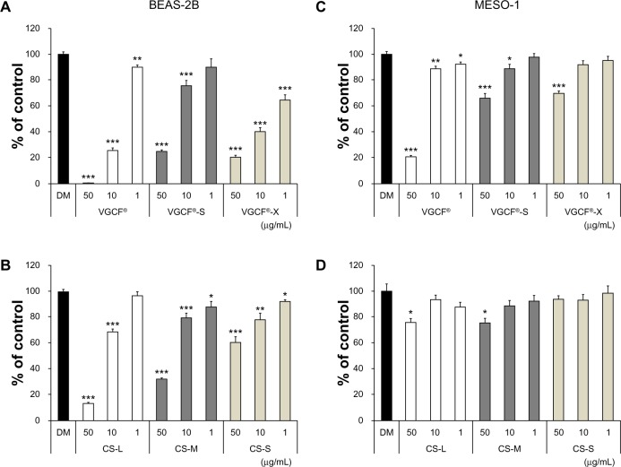
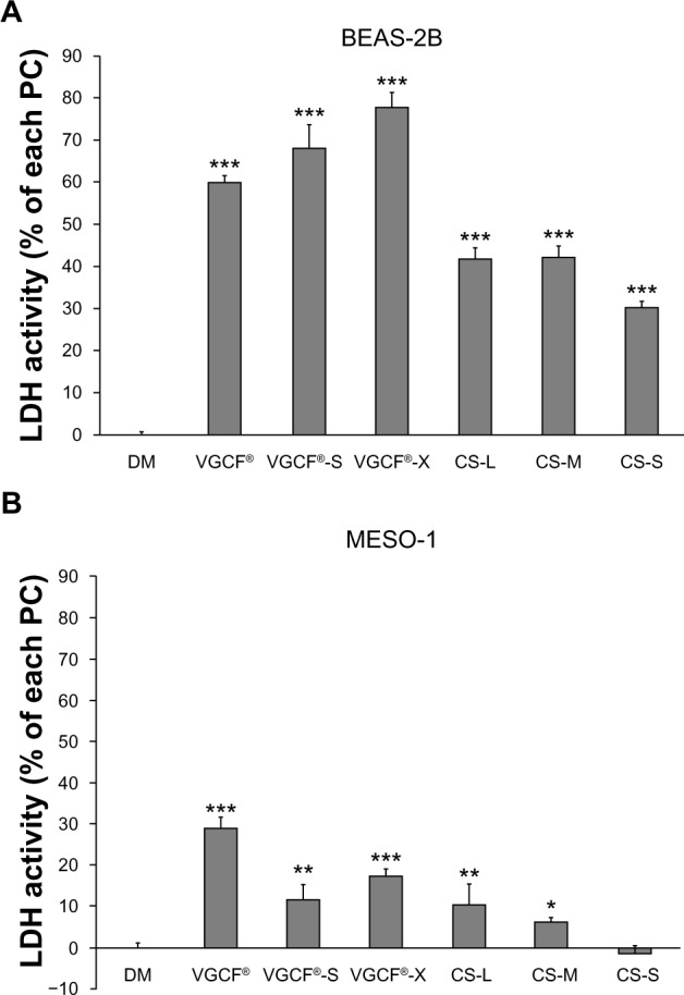
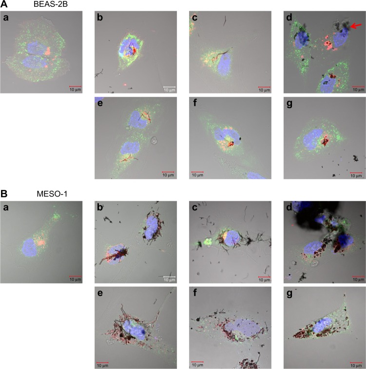
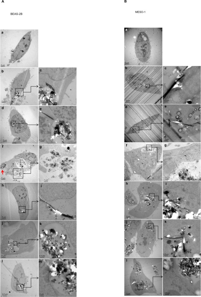
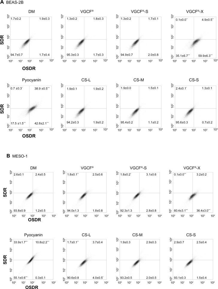
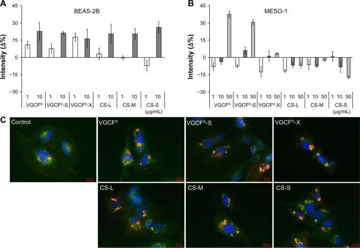
This study aimed to investigate the influence of the shape and size of multi-walled carbon nanotubes (MWCNTs) and cup-stacked carbon nanotubes (CSCNTs) on biological responses in vitro. Three types of MWCNTs - VGCF(®)-X, VGCF(®)-S, and VGCF(®) (vapor grown carbon fibers; with diameters of 15, 80, and 150 nm, respectively) - and three CSCNTs of different lengths (CS-L, 20-80 μm; CS-S, 0.5-20 μm; and CS-M, of intermediate length) were tested. Human bronchial epithelial (BEAS-2B) and malignant pleural mesothelioma cells were exposed to the CNTs (1-50 μg/mL), and cell viability, permeability, uptake, total reactive oxygen species/superoxide production, and intracellular acidity were measured. CSCNTs were less toxic than MWCNTs in both cell types over a 24-hour exposure period. The cytotoxicity of endocytosed MWCNTs varied according to cell type/size, while that of CSCNTs depended on tube length irrespective of cell type. CNT diameter and length influenced cell aggregation and injury extent. Intracellular acidity increased independently of lysosomal activity along with the number of vacuoles in BEAS-2B cells exposed for 24 hours to either CNT (concentration, 10 μg/mL). However, total reactive oxygen species/superoxide generation did not contribute to cytotoxicity. The results demonstrate that CSCNTs could be suitable for biological applications and that CNT shape and size can have differential effects depending on cell type, which can be exploited in the development of highly specialized, biocompatible CNTs.

Keywords: cup-stacked carbon nanotube; cytotoxicity; in vitro; intracellular acidity; multi-walled carbon nanotube.


Figures

Figure 1
Figure 1
Cell viability upon exposure to CNTs. Notes: Cells were exposed to the indicated concentrations of CNT for 24 hours. BEAS-2B cells exposed to (A) MWCNTs (VGCF®-X, VGCF®-S, and VGCF®) and (B) CSCNTs (CS-L, CS-S, and CS-M). MESO-1 cells exposed to (C) MWCNTs and (D) CSCNTs. DM (0.001% gelatin) served as the control, and data are expressed as mean ± standard error (n=6). *P<0.05; **P<0.01; ***P<0.001. MWCNTs were provided by Showa Denko KK (Tokyo, Japan); CSCNTs were provided by GSI Creos (Tokyo, Japan); VGCF, vapor grown carbon fibers; CS-L, CSCNT of length 20–80 μm; CS-S, CSCNT of length 0.5–20 μm; CS-M, CSCNT of intermediate length. Abbreviations: CNT, carbon nanotube; CSCNT, cup-stacked CNT; DM, dispersant medium; MWCNT, multi-walled CNT; VGCF, vapor grown carbon fibers; CS-L, CSCNT of length 20–80 μm; CS-S, CSCNT of length 0.5–20 μm; CS-M, CSCNT of intermediate length.
Figure 2
Figure 2
Plasma membrane permeability in cells exposed to CNTs. Notes: Cells were exposed to 10 μg/mL CNT for 24 hours. (A) BEAS-2B and (B) MESO-1 cells exposed to MWCNTs or CSCNTs. The LDH activity was calculated by the formula ([experimental value − DM value]/[PC value − DM value]) ×10 × 100 (%). PC is 0.01% Triton X-100; DM is 0.001% gelatin. Data are compared to the control (DM) and expressed as mean ± standard error (n=3). *P<0.05; **P<0.01; ***P<0.001. MWCNTs were provided by Showa Denko KK (Tokyo, Japan); CSCNTs were provided by GSI Creos (Tokyo, Japan). Abbreviations: CNT, carbon nanotube; CSCNT, cup-stacked CNT; DM, dispersant medium; LDH, lactate dehydrogenase; MWCNT, multi-walled CNT; PC, positive control; VGCF, vapor grown carbon fibers; CS-L, CSCNT of length 20–80 μm; CS-S, CSCNT of length 0.5–20 μm; CS-M, CSCNT of intermediate length.
Figure 3
Figure 3
Live cells imaged by differential interference contrast optics after incubation with CellLight® Lysosomes-RFP and Early Endosomes-GFP (Life Technologies, Carlsbad, CA, USA) and bisbenzimide H33342 fluorochrome trihydrochloride for nuclear staining. Notes: (A) BEAS-2B cells were exposed to 1 μg/mL MWCNT (VGCF) or CSCNT (CS) for 24 hours. (a) DM (control); (b) VGCF®; (c) VGCF®-S; (d) VGCF®-X; (e) CS-L; (f) CS-M; and (g) CS-S. Red arrow indicates VGCF-X agglomerates, which were not taken up by BEAS-2B cells, nor did they fully penetrate the cell membrane. Scale bar =10 μm. (B) MESO-1 cells were exposed to 10 μg/mL MWCNT (VGCF) or CSCNT (CS) for 24 hours. (a) DM (control); (b) VGCF; (c) VGCF-S; (d) VGCF-X; (e) CS-L; (f) CS-M; and (g) CS-S. Scale bar =10 μm. MWCNTs were provided by Showa Denko KK (Tokyo, Japan); CSCNTs were provided by GSI Creos (Tokyo, Japan). Abbreviations: CSCNT, cup-stacked carbon nanotube; DM, dispersant medium; MWCNT, multi-walled carbon nanotube; CS-L, CSCNT of length 20–80 μm; CS-S, CSCNT of length 0.5–20 μm; CS-M, CSCNT of intermediate length.
Figure 4
Figure 4
Transmission electron micrographs of BEAS-2B and MESO-1 cells exposed to CNTs. Notes: (A) BEAS-2B cells were exposed to 1 μg/mL MWCNT (VGCF) or CSCNT (CS) for 24 hours. (a) DM (control); (b and c) VGCF®; (d and e) VGCF®-S; (f and g) VGCF®-X; (h and i) CS-L; (j and k) CS-M; and (l and m) CS-S. Red arrow indicates VGCF-X agglomerates, which were not taken up by BEAS-2B cells, nor did they fully penetrate the cell membrane. (B) MESO-1 cells were exposed to 10 μg/mL MWCNT or CSCNT for 24 hours. (a) DM (control); (b and c) VGCF; (d and e) VGCF-S; (f and g) VGCF-X; (h and i) CS-L; (j and k) CS-M; and (l and m) CS-S. MWCNTs were provided by Showa Denko KK (Tokyo, Japan); CSCNTs were provided by GSI Creos (Tokyo, Japan). Abbreviations: CNT, carbon nanotube; CSCNT, cup-stacked carbon nanotube; DM, dispersant medium; MWCNT, multi-walled carbon nanotube; CS-L, CSCNT of length 20–80 μm; CS-S, CSCNT of length 0.5–20 μm; CS-M, CSCNT of intermediate length; VGCF, vapor grown carbon fibers.
Figure 5
Figure 5
Total reactive oxygen species production upon exposure to CNTs. Notes: (A) BEAS-2B cells were exposed to 1 μg/mL MWCNT (VGCF®, VGCF®-S, and VGCF®-X) or CSCNT (CS-L, CS-M, and CS-S) for 1 hour. (B) MESO-1 cells were exposed to 10 μg/mL MWCNT (VGCF, VGCF-S, and VGCF-X) or CSCNT (CS-L, CS-M, and CS-S) for 1 hour. Pyocyanin (100 μM) was used to induce the production of reactive oxygen species. Data are expressed as mean ± standard error (n=3). *P<0.05; **P<0.01; ***P<0.001. MWCNTs were provided by Showa Denko KK (Tokyo, Japan); CSCNTs were provided by GSI Creos (Tokyo, Japan). Abbreviations: CNT, carbon nanotube; CSCNT, cup-stacked carbon nanotube; DM, dispersant medium; MWCNT, multi-walled carbon nanotube; OSDR, oxidative stress detection reagent; SDR, superoxide detection reagent; CS-L, CSCNT of length 20–80 μm; CS-S, CSCNT of length 0.5–20 μm; CS-M, CSCNT of intermediate length; VGCF, vapor grown carbon fibers.
Figure 6
Figure 6
Intracellular acidification upon exposure to CNTs. Notes: (A) BEAS-2B cells were exposed to 1 and 10 μg/mL of MWCNT (VGCF®-X, VGCF®-S, and VGCF®) or CSCNT (CS-L, CS-S, and CS-M) for 24 hours. (B) MESO-1 cells were exposed to 1, 10, and 50 μg/mL MWCNT or CSCNT for 24 hours, incubated with an acidotropic probe (LysoSensor™; Life Technologies, Carlsbad, CA, USA), and analyzed by flow cytometry (10,000 cells). The LysoSensor intensity (%) was calculated as follows: ([FL1 channel intensity of cells exposed to CNTs − FL1 channel intensity of untreated cells]/[FL1 channel intensity of cells exposed to DM − FL1 channel intensity of untreated cells]) ×100%. DM is 0.001% gelatin. CNT inhibition intensity (%) was calculated as follows: ([FL1 channel intensity of CNT blank − FL1 channel intensity of untreated cells]/[FL1 channel intensity of DM blank − FL1 channel intensity of untreated cells]) ×100%. The intensity change was determined as follows: intensity (Δ%) = LysoSensor intensity (%) − CNT inhibition intensity (%). Data are expressed as mean ± standard error (n=4). (C) BEAS-2B cells were exposed to 10 μg/mL MWCNT (VGCF, VGCF-S, and VGCF-X) or CSCNT (CS-L, CS-M, and CS-S) for 24 hours and visualized by staining with LysoSensor dye (green), CellLight® Lysosomes-RFP (red [Life Technologies]), and bisbenzimide H33342 fluorochrome trihydrochloride (blue [Nacalai Tesque, Kyoto, Japan]) for nuclei. Scale bar =20 μm. MWCNTs were provided by Showa Denko KK (Tokyo, Japan); CSCNTs were provided by GSI Creos (Tokyo, Japan). Abbreviations: CNT, carbon nanotube; CSCNT, cup-stacked carbon nanotube; DM, dispersant medium; MWCNT, multi-walled carbon nanotube; CS-L, CSCNT of length 20–80 μm; CS-S, CSCNT of length 0.5–20 μm; CS-M, CSCNT of intermediate length; VGCF, vapor grown carbon fibers.

References

    1. Saito N, Usui Y, Aoki K, et al. Carbon nanotubes: biomaterial applications. Chem Soc Rev. 2009;38(7):1897–1903. - PubMed
    1. Li X, Fan Y, Watari F. Current investigations into carbon nanotubes for biomedical application. Biomed Mater. 2010;5(2):22001. - PubMed
    1. Adams D, Williams DF. The response of bone to carbon – carbon composites. Biomaterials. 1984;5(2):59–64. - PubMed
    1. Bokros JC. Carbon biomedical devices. Carbon. 1977;15(6):353–371.
    1. Poland CA, Duffin R, Kinloch I, et al. Carbon nanotubes introduced into the abdominal cavity of mice show asbestos-like pathogenicity in a pilot study. Nat Nanotechnol. 2008;3(7):423–428. - PubMed

Publication types

LinkOut - more resources