

DDIT3 Expression in Liposarcoma Development
- PMID: 24790523
- PMCID: PMC3984824
- DOI: 10.1155/2014/954671
DDIT3 Expression in Liposarcoma Development
Abstract
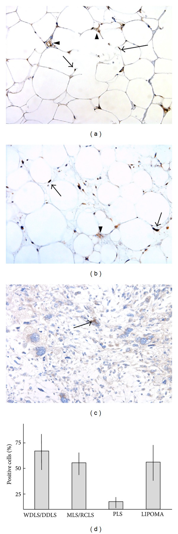
Liposarcomas are mesenchymal tumors containing variable numbers of lipoblasts or adipocytes. The most common entities, well differentiated/dedifferentiated liposarcoma (WDLS/DDLS) and myxoid/round cell liposarcoma (MLS/RCLS), are both characterized by genetic rearrangements that affect the expression of the transcription factor DDIT3. DDIT3 induces liposarcoma morphology when ectopically expressed in a human fibrosarcoma. The role of DDIT3 in lipomatous tumors is, however, unclear. We have analyzed the expression of DDIT3 in 37 cases of liposarcoma (WDLS/DDLS n = 10, MLS/RCLS n = 16, and pleomorphic liposarcomas (PLS) n = 11) and 11 cases of common benign lipomas. Major cell subpopulations of WDLS/DDLS and MLS/RCLS tumors were found to express DDIT3 or the derived fusion protein, whereas PLS cases showed only a few positive cells. The lipomas contained large subpopulations expressing DDIT3. No correlation between numbers of DDIT3 expressing cells and numbers of lipoblasts/adipocytes was found. In vitro adipogenic treatment of two DDIT3 expressing cell lines induced lipid accumulation in small subpopulations only. Our results suggest a dual, promoting and limiting, role for DDIT3 in the formation of lipoblasts and liposarcoma morphology.
Figures

Similar articles
-
The myxoid/round cell liposarcoma fusion oncogene FUS-DDIT3 and the normal DDIT3 induce a liposarcoma phenotype in transfected human fibrosarcoma cells.Am J Pathol. 2006 May;168(5):1642-53. doi: 10.2353/ajpath.2006.050872. Am J Pathol. 2006. PMID: 16651630 Free PMC article.
-
Nuclear expression of FLT1 and its ligand PGF in FUS-DDIT3 carrying myxoid liposarcomas suggests the existence of an intracrine signaling loop.BMC Cancer. 2010 Jun 1;10:249. doi: 10.1186/1471-2407-10-249. BMC Cancer. 2010. PMID: 20515481 Free PMC article.
-
Lipomatous tumors.Monogr Pathol. 1996;38:207-39. Monogr Pathol. 1996. PMID: 8744279 Review.
-
DDIT3 Immunohistochemistry Is a Useful Tool for the Diagnosis of Myxoid Liposarcoma.Am J Surg Pathol. 2021 Feb 1;45(2):230-239. doi: 10.1097/PAS.0000000000001564. Am J Surg Pathol. 2021. PMID: 32815829 Free PMC article.
-
Liposarcomas of the mediastinum.Mediastinum. 2020 Sep 30;4:27. doi: 10.21037/med-20-42. eCollection 2020. Mediastinum. 2020. PMID: 35118295 Free PMC article. Review.
Cited by
-
Axitinib Has Antiangiogenic and Antitumorigenic Activity in Myxoid Liposarcoma.Sarcoma. 2016;2016:3484673. doi: 10.1155/2016/3484673. Epub 2016 Oct 16. Sarcoma. 2016. PMID: 27822137 Free PMC article.
-
Linc00423 as a tumor suppressor in retroperitoneal liposarcoma via activing MAPK signaling pathway through destabilizing of NFATC3.Cell Death Dis. 2019 Jun 3;10(6):430. doi: 10.1038/s41419-019-1658-2. Cell Death Dis. 2019. PMID: 31160581 Free PMC article.
-
FUS::DDIT3 Fusion Protein in the Development of Myxoid Liposarcoma and Possible Implications for Therapy.Biomolecules. 2024 Oct 14;14(10):1297. doi: 10.3390/biom14101297. Biomolecules. 2024. PMID: 39456230 Free PMC article. Review.
-
Analysis of expression profile data identifies key genes and pathways in hepatocellular carcinoma.Oncol Lett. 2018 Feb;15(2):2625-2630. doi: 10.3892/ol.2017.7534. Epub 2017 Dec 6. Oncol Lett. 2018. PMID: 29434983 Free PMC article.
-
Well-Differentiated Liposarcoma of the Hypopharynx Exhibiting Myxoid Liposarcoma-like Morphology with MDM2 and DDIT3 Co-Amplification.Head Neck Pathol. 2022 Mar;16(1):288-293. doi: 10.1007/s12105-021-01341-5. Epub 2021 Jun 4. Head Neck Pathol. 2022. PMID: 34089125 Free PMC article.
References
-
- Fletcher CDM, Krishnan Unni K, Mertens F. Tumors of Soft Tissue and Bone. IARC Press; 2000.
-
- Pedeutour F, Forus A, Berner JM, et al. Structure of the supernumerary ring and giant rod chromosomes in adipose tissue tumors. Genes, Chromosomes & Cancer. 1999;24(1):30–41. - PubMed
-
- Pedeutour F, Suijkerbuijk RF, Forus A, et al. Complex composition and co-amplification of SAS and MDM2 in ring and giant rod marker chromosomes in well-differentiated liposarcoma. Genes, Chromosomes & Cancer. 1994;10(2):85–94. - PubMed
-
- Persson F, Olofsson A, Sjögren H, et al. Characterization of the 12q amplicons by high-resolution, oligonucleotide array CGH and expression analyses of a novel liposarcoma cell line. Cancer Letters. 2008;260(1-2):37–47. - PubMed
-
- Crozat A, Aman P, Mandahl N, Ron D. Fusion of CHOP to a novel RNA-binding protein in human myxoid liposarcoma. Nature. 1993;363(6430):640–644. - PubMed
LinkOut - more resources
Full Text Sources
Other Literature Sources
Research Materials
