. 2014:2014:537865.
doi: 10.1155/2014/537865. Epub 2014 Mar 23.

PPARG in Human Adipogenesis: Differential Contribution of Canonical Transcripts and Dominant Negative Isoforms

Affiliations

PPARG in Human Adipogenesis: Differential Contribution of Canonical Transcripts and Dominant Negative Isoforms

M Aprile et al. PPAR Res. 2014.

Abstract

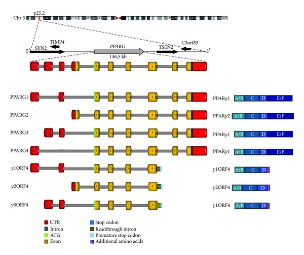
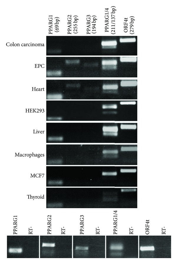
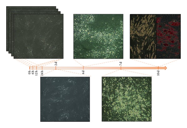
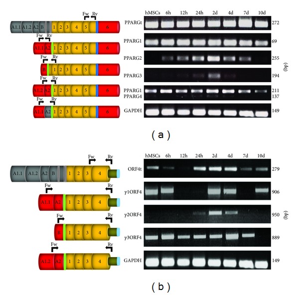
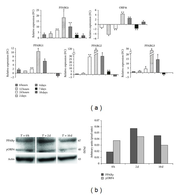
The nuclear receptor PPAR γ is a key regulator of adipogenesis, and alterations of its function are associated with different pathological processes related to metabolic syndrome. We recently identified two PPARG transcripts encoding dominant negative PPAR γ isoforms. The existence of different PPARG variants suggests that alternative splicing is crucial to modulate PPAR γ function, underlying some underestimated aspects of its regulation. Here we investigate PPARG expression in different tissues and cells affected in metabolic syndrome and, in particular, during adipocyte differentiation of human mesenchymal stem cells. We defined the transcript-specific expression pattern of PPARG variants encoding both canonical and dominant negative isoforms and identified a novel PPARG transcript, γ 1ORF4. Our analysis indicated that, during adipogenesis, the transcription of alternative PPARG variants is regulated in a time-specific manner through differential usage of distinct promoters. In addition, our analysis describes-for the first time-the differential contribution of three ORF4 variants to this process, suggesting a still unexplored role for these dominant negative isoforms during adipogenesis. Therefore, our results highlight crucial aspects of PPARG regulation, suggesting the need of further investigation to rule out the differential impact of all PPARG transcripts in both physiologic and pathologic conditions, such as metabolism-related disorders.


Figures

Figure 1
Figure 1
Schematic representation of human PPARG gene, transcripts, and protein isoforms. In the upper part the genomic localization of PPARG gene is indicated, with chromosome indication, cytogenetic band, and surrounding genes. Below is depicted the exon/intron structure of PPARG gene with transcribed splicing variants. Transcripts encoding both the canonical and dominant negative proteins are illustrated in the left panel. The right panel shows a schematic representation of the encoded proteins with the functional domains.
Figure 2
Figure 2
Expression pattern of PPARG variants in tissues and cells affected in the metabolic syndrome. For each PPARG transcript, specific primer pairs were used for PCR reactions. Given the similarity between PPARG1/4 5′UTRs primers amplifies both variants (distinguishable as PCR products of different sizes). “ORF4t” indicates the entire pool of ORF4 transcripts. Amplicons' sizes are shown (in bp) below transcripts' names. On the bottom panel, negative PCR controls are shown for each primer pair.
Figure 3
Figure 3
Phenotypic characteristics of undifferentiated, differentiating, and differentiated hMSCs (h = hours; d = days). Adipocyte differentiation was determined at 10 days from adipogenesis induction by Oil Red O staining of lipids vacuoles, as shown.
Figure 4
Figure 4
Transcript-specific RT-PCR assays for PPARG canonical transcripts (panel (a)) and ORF4 variants (panel (b)) at different time points of the adipogenesis (indicated on the top). On the left, the different PPARG transcripts are schematically shown; on the right the related PCR amplicons and their sizes (in bp) are illustrated. “PPARGt” and “ORF4t” indicate the entire pool of canonical PPARG and ORF4 transcripts, respectively. Transcript-specific exons are shown in grey and common exons are coloured.Black arrows indicate the specific primer pairs used in this analysis (Fw, forward; Rv, reverse). GAPDH was used as internal control.
Figure 5
Figure 5
For each analyzed PPARG variant, bar graphs in the Panel (a) indicate the relative expression levels at different time points after in vitro adipocyte differentiation. For each assay, expression is normalized for reference samples (time point at 0 or 6 hours) using GAPDH as housekeeping gene. Data are reported as mean values, and error bars are also reported. P values <0.05 are considered statistically significant and indicated by an asterisk. Double asterisks indicate P values <0.001. In panel (b), total cell lysates of hMSC at day 0, day 2, and day 10 by differentiation induction blotted with anti-PPARγ antibody are shown. To ensure equal protein transfer, membranes were blotted with antiactin antibody. Bar graph indicates the pixel intensity ratio between PPARγ isoforms and actin protein levels, reported as arbitrary units over basal (day 0).

References

    1. Willson TM, Brown PJ, Sternbach DD, Henke BR. The PPARs: from orphan receptors to drug discovery. Journal of Medicinal Chemistry. 2000;43(4):527–550. - PubMed
    1. Cho MC, Lee K, Paik SG, Yoon DY. Peroxisome proliferators-activated receptor (PPAR) modulators and metabolic disorders. PPAR Research. 2008;2008:14 pages.679137 - PMC - PubMed
    1. Costa V, Gallo MA, Letizia F, Aprile M, Casamassimi A, Ciccodicola A. PPARG: gene expression regulation and next-generation sequencing for unsolved issues. PPAR Research. 2010;2010:17 pages.409168 - PMC - PubMed
    1. Rosen ED, Spiegelman BM. PPARγ: a nuclear regulator of metabolism, differentiation, and cell growth. Journal of Biological Chemistry. 2001;276(41):37731–37734. - PubMed
    1. Ahmed W, Ziouzenkova O, Brown J, et al. PPARs and their metabolic modulation: new mechanisms for transcriptional regulation? Journal of Internal Medicine. 2007;262(2):184–198. - PubMed

LinkOut - more resources