

Unifocal orofacial granulomatosis in retromolar mucosa: surgical treatment with Er,Cr:YSGG laser
- PMID: 24790722
- PMCID: PMC4002352
- DOI: 10.4317/jced.51301
Unifocal orofacial granulomatosis in retromolar mucosa: surgical treatment with Er,Cr:YSGG laser
Abstract
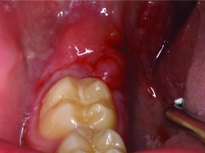
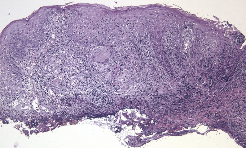
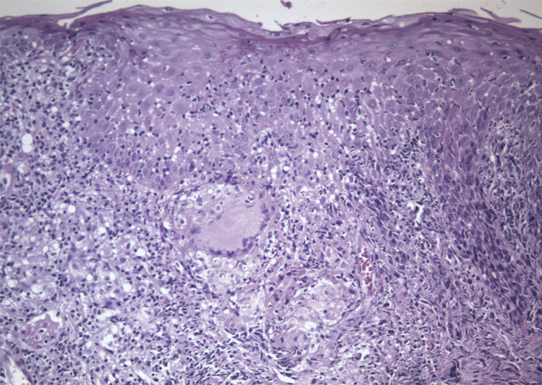
Orofacial granulomatosis is defined by permanent or recurrent swelling of orofacial tissues with different multiform and multifocal clinical patterns. An 11-year old boy presented with a 2-month history of mucosa enlargement. Intraoral examination revealed an erythematous, polylobulated, exophytic lesion with a smooth surface located in retromolar mucosa, non-tender and non-infiltratated to palpation. The diagnosis was inflammatory lesion compatible with pyogenic granuloma and laser excision was decided. Haematological parameters were within normal range, as well as chest Xrays. These findings lead to a diagnosis of non-symptomatic orofacial granulomatosis, whose early diagnosis can minimize the impact of systemic-related disorders, like Chron's disease. Key words:Laser, orofacial granulomatosis, childhood, oral lesions, diagnosis.
Conflict of interest statement
Figures
Similar articles
-
Idiopathic orofacial granulomatosis - a diagnostic and treatment challenge.J Clin Diagn Res. 2014 Nov;8(11):ZD07-10. doi: 10.7860/JCDR/2014/10047.4961. Epub 2014 Nov 20. J Clin Diagn Res. 2014. PMID: 25584331 Free PMC article.
-
Orofacial Granulomatosis in Children can be the Initial Manifestation of Systemic Disease: A Presentation of Two Cases.Dermatol Reports. 2014 May 12;6(1):5039. doi: 10.4081/dr.2014.5039. eCollection 2014 Feb 17. Dermatol Reports. 2014. PMID: 25386323 Free PMC article.
-
Orofacial granulomatosis affecting lip and gingiva in a 15-year-old patient: A rare case report.Contemp Clin Dent. 2015 Mar;6(Suppl 1):S94-6. doi: 10.4103/0976-237X.152958. Contemp Clin Dent. 2015. PMID: 25821385 Free PMC article.
-
Orofacial Granulomatosis.Dermatol Clin. 2015 Jul;33(3):433-46. doi: 10.1016/j.det.2015.03.008. Epub 2015 May 6. Dermatol Clin. 2015. PMID: 26143423 Review.
-
Erythematous and Vascular Oral Mucosal Lesions: A Clinicopathologic Review of Red Entities.Head Neck Pathol. 2019 Mar;13(1):4-15. doi: 10.1007/s12105-019-01002-8. Epub 2019 Jan 29. Head Neck Pathol. 2019. PMID: 30693460 Free PMC article. Review.
Cited by
-
Evaluation of the efficacy of Er,Cr:YSGG laser in Treating oral benign soft tissue lesions.J Dent Res Dent Clin Dent Prospects. 2024 Fall;18(4):291-296. doi: 10.34172/joddd.40905. Epub 2024 Dec 14. J Dent Res Dent Clin Dent Prospects. 2024. PMID: 39895680 Free PMC article.
-
Uncommon inflammatory swelling of the lips: orofacial granulomatosis.BMJ Case Rep. 2016 Jan 12;2016:bcr2015211860. doi: 10.1136/bcr-2015-211860. BMJ Case Rep. 2016. PMID: 26759437 Free PMC article.
References
-
- Leão JC, Hodgson T, Scully C, Porter S. Review article: orofacial granulomatosis. Aliment Pharmacol Ther. 2004;20:1019–27. - PubMed
-
- Hegarty A, Hodgson T, Porter S. Thalidomide for the treatment of recalcitrant oral Crohn´s disease and orofacial granulomatosis. Oral Surg Oral Med Oral Pathol Oral Radiol Endod. 2003;95:576–85. - PubMed
-
- Grave B, McCullough M, Wiesenfeld D. Orofacial granulomatosis – a 20-year review. Oral Dis. 2009;15:46–51. - PubMed
-
- Lourenço SV, Lobo AZ, Boggio P, Fezzi F, Sebastião A, Nico MM. Gingival manifestations of orofacial granulomatosis. Arch Dermatol. 2008;12:1627–30. - PubMed
-
- Fitzpatrick L, Healy CM, McCartan BE, Flint SR, McCreary CE, Rogers S. Patch testing for food-associated allergies in orofacial granulomatosis. J Oral Pathol Med. 2011;40:10–3. - PubMed
Publication types
LinkOut - more resources
Full Text Sources
Other Literature Sources