. 2013 Aug 22;1(1):10.

Multiple Functional Motifs Are Required for the Tumor Suppressor Activity of a Constitutively-Active ErbB4 Mutant

Affiliations

Multiple Functional Motifs Are Required for the Tumor Suppressor Activity of a Constitutively-Active ErbB4 Mutant

Richard M Gallo et al. J Cancer Res Ther Oncol. .

Abstract

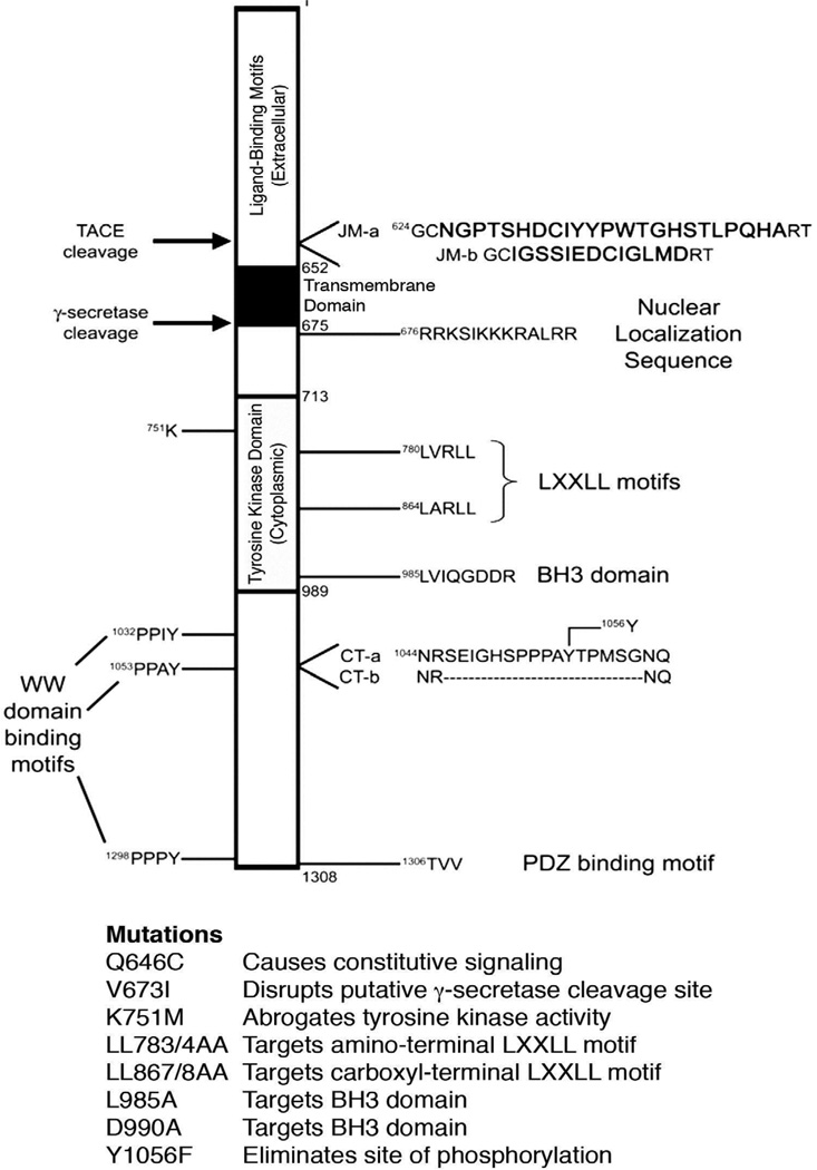
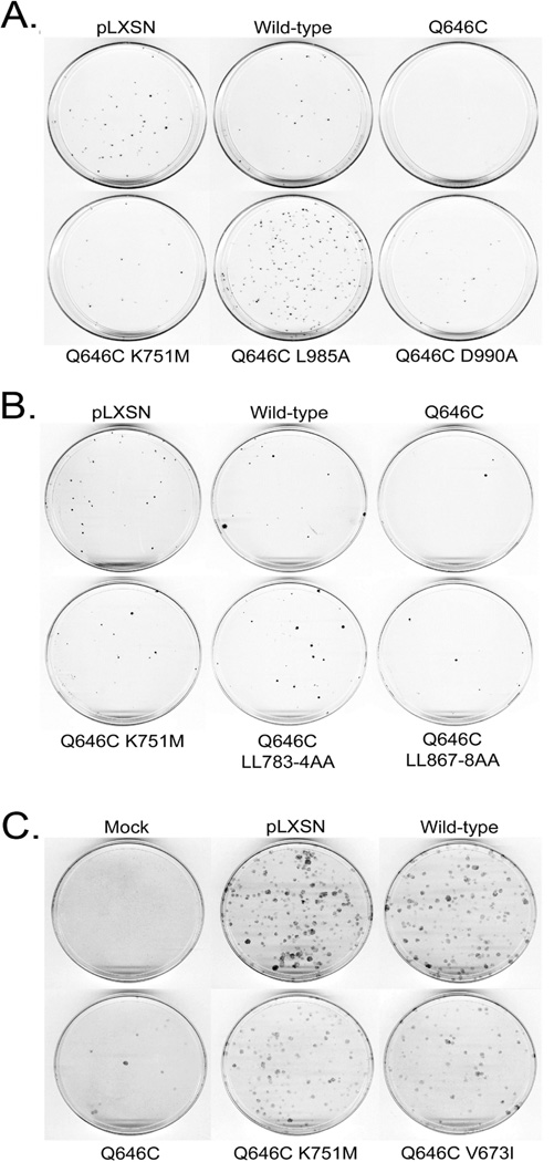
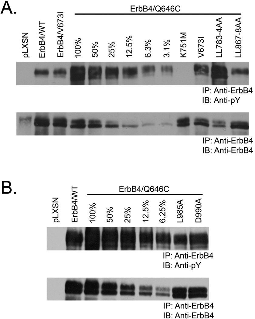
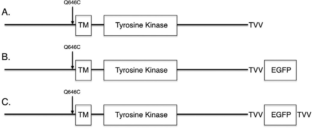
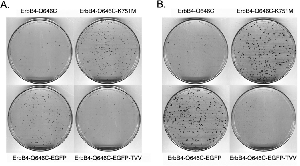
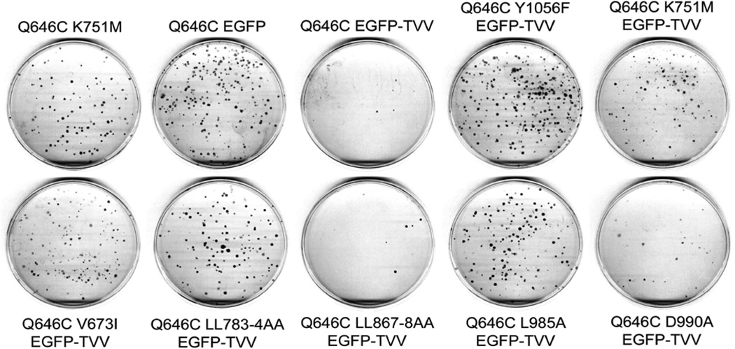
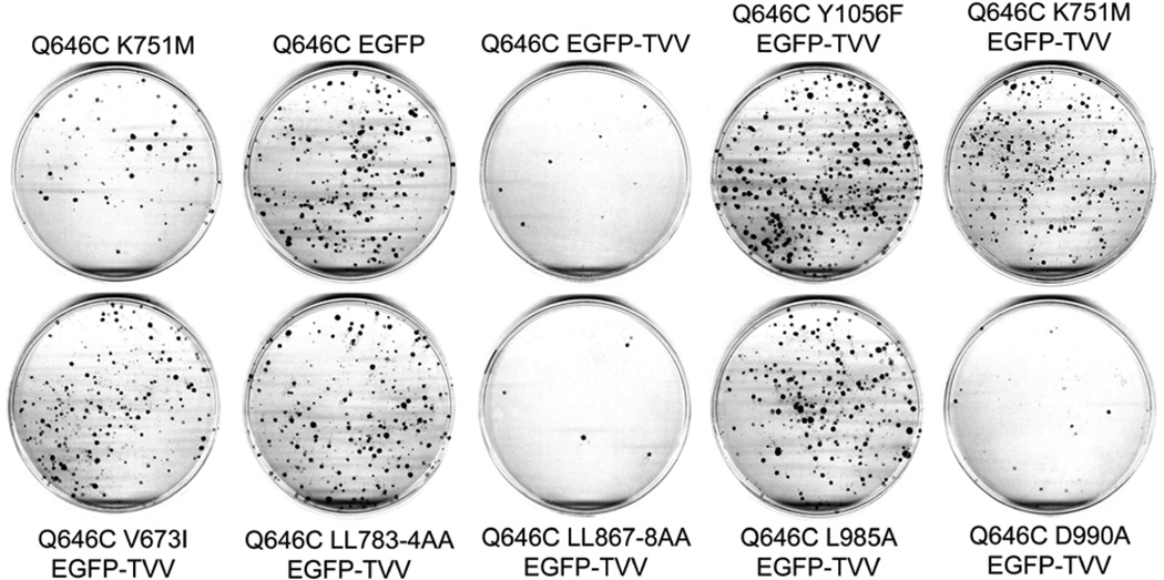
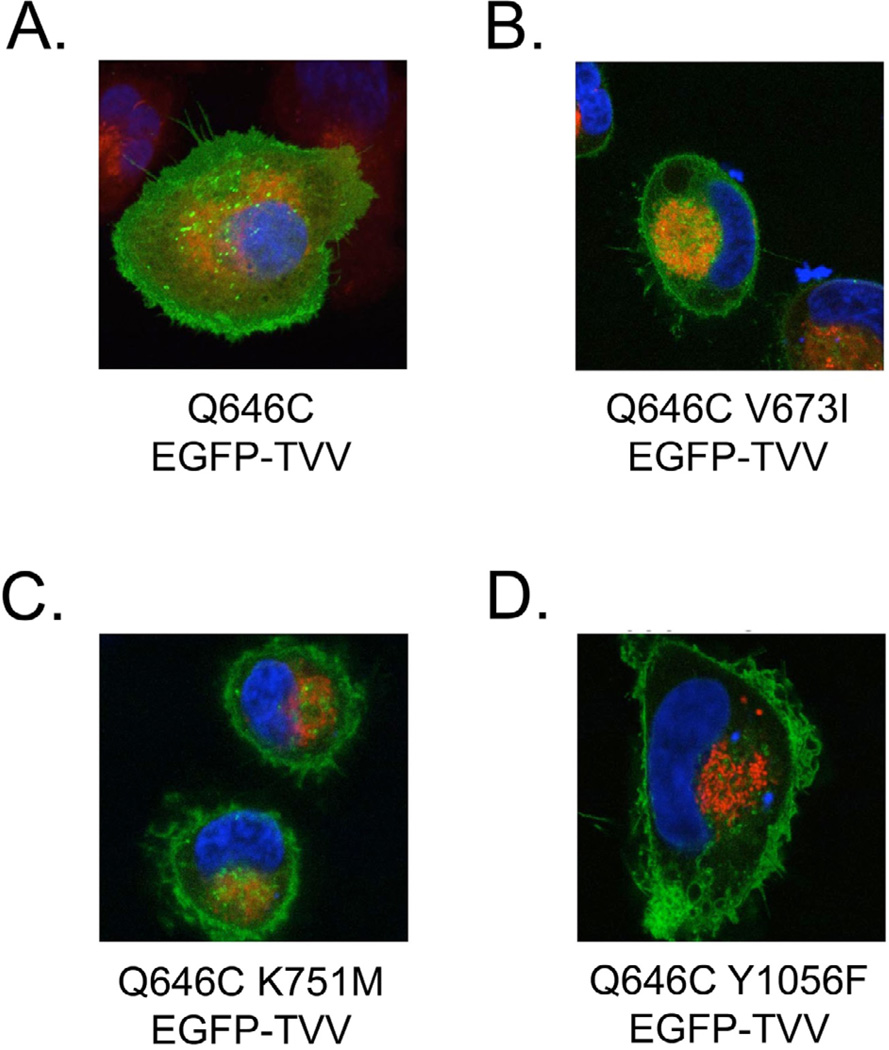
ErbB4 (HER4) is a member of the ErbB family of receptor tyrosine kinases, which includes the Epidermal Growth Factor Receptor (EGFR/ErbB1), ErbB2 (HER2/Neu), and ErbB3 (HER3). Mounting evidence indicates that ErbB4, unlike EGFR or ErbB2, functions as a tumor suppressor in many human malignancies. Previous analyses of the constitutively-dimerized and -active ErbB4 Q646C mutant indicate that ErbB4 kinase activity and phosphorylation of ErbB4 Tyr1056 are both required for the tumor suppressor activity of this mutant in human breast, prostate, and pancreatic cancer cell lines. However, the cytoplasmic region of ErbB4 possesses additional putative functional motifs, and the contributions of these functional motifs to ErbB4 tumor suppressor activity have been largely underexplored. Here we demonstrate that ErbB4 BH3 and LXXLL motifs, which are thought to mediate interactions with Bcl family proteins and steroid hormone receptors, respectively, are required for the tumor suppressor activity of the ErbB4 Q646C mutant. Furthermore, abrogation of the site of ErbB4 cleavage by gamma-secretase also disrupts the tumor suppressor activity of the ErbB4 Q646C mutant. This last result suggests that ErbB4 cleavage and subcellular trafficking of the ErbB4 cytoplasmic domain may be required for the tumor suppressor activity of the ErbB4 Q646C mutant. Indeed, here we demonstrate that mutants that disrupt ErbB4 kinase activity, ErbB4 phosphorylation at Tyr1056, or ErbB4 cleavage by gamma-secretase also disrupt ErbB4 trafficking away from the plasma membrane and to the cytoplasm. This supports a model for ErbB4 function in which ErbB4 tumor suppressor activity is dependent on ErbB4 trafficking away from the plasma membrane and to the cytoplasm, mitochondria, and/or the nucleus.

Keywords: ErbB4/HER4; Protein Trafficking; Signal Transduction; Tumor Suppressor.


Figures

Figure 1
Figure 1. ErbB4 Possesses Multiple Functional Motifs and Mutations Have Been Engineered to Target These Motifs
The organization of ErbB4 is as indicated in this schematic. The extracellular ligand-binding motifs reside in the amino-terminal region upstream of amino acid residue 651. The single-pass transmembrane domain consists of amino acid residues 652–675. The cytoplasmic tyrosine kinase domain consists of amino acid residues 713–989. The majority of cytoplasmic sites of tyrosine phosphorylation reside in amino acid residues 990–1308, most notably Tyr1056. Additional putative functional motifs include a TACE cleavage site, a gamma-secretase cleavage site, two LXXLL (steroid hormone receptor binding) motifs, a BH3 domain, three WW domain binding motifs, and a PDZ domain binding motif. Mutations that disrupt these motifs are noted. Finally, note the two locations of alternative transcriptional splicing, resulting in a total of four different splicing isoforms.
Figure 2
Figure 2. K751M, V673I, LL783/4AA, and L985A mutations profoundly disrupt the tumor suppressor activity of the constitutively-activeErbB4 Q646C construct in MCF7 and MCF10A cell lines
(a,b) MCF7 cells were infected with recombinant retroviruses based on the pLXSN vector as indicated. Infected cells were selected using G418 and colonies of infected, drug-resistant cells were stained using Giemsa and photographed. Images are representative of four independent experiments. The number of colonies was counted and the effects of the various constructs on clonogenic proliferation of MCF7 cells were analyzed as indicated elsewhere and reported in Table 1. (c) MCF10A cells were infected with recombinant retroviruses based on the pLXSN vector as indicated. Infected cells were selected using G418 and colonies of infected, drug-resistant cells were stained using Giemsa and photographed. Images are representative of at least three independent experiments. The number of colonies was counted and the effects of the various constructs on clonogenic proliferation of MCF10A cells were analyzed as indicated elsewhere and reported in Table 2.
Figure 3
Figure 3. The V673I, LL783/4AA, and L985A mutations do not markedly alter the expression or tyrosine phosphorylation of the constitutively-active ErbB4 Q646C construct
The expression and tyrosine phosphorylation of ErbB4 mutants expressed in Ψ2 cell lines generated in the course of the experiments described in Figure 2 were analyzed by ErbB4 immunoprecipitation of Ψ2 cell lysates and ErbB4 or anti-phosphotyrosine immunoblotting as described previously [40, 41]. These experiments were performed using 1 mg of lysate unless otherwise noted (100% indicates that 1 mg of lysate was used, 50% indicates that 0.5 mg of lysate was used, 25% indicates that 0.25 mg of lysate was used, and so on). Images are representative of three independent experiments.
Figure 4
Figure 4. An Enhanced Green Fluorescent Protein (EGFP) tag and the amino acid sequence Thr-Val-Val (TVV) have been added to the carboxyl terminus of the ErbB4 Q646C mutant
Standard subcloning techniques were used to move the EGFP sequence from the ErbB4-EGFP construct [10] to the ErbB4 Q646C construct [41], thereby generating the ErbB4 Q646C EGFP construct. Standard site-directed mutagenesis techniques were used to add the Thr-Val-Val (TVV) sequence to the extreme carboxyl terminus of the ErbB4 Q646C EGFP construct, thereby generating the ErbB4 Q646C EGFP-TVV construct.
Figure 5
Figure 5. Adding a carboxyl-terminal Enhanced Green Fluorescent Protein (EGFP) tag to the constitutively-active ErbB4 Q646C mutant disrupts its tumor suppressor activity, but this deficit is rescued by adding a carboxyl-terminal Thr-Val-Val (TVV) sequence
(a) PC-3 cells were infected with recombinant retroviruses based on the pLXSN-ErbB4 vector as indicated. Infected cells were selected using G418 and colonies of infected, drug-resistant cells were stained using Giemsa and photographed. Images are representative of three independent experiments. The number of colonies was counted and the effects of the various constructs on clonogenic proliferation of PC-3 cells were analyzed as indicated elsewhere and reported in Table 3. (b) DU-145 cells were infected with recombinant retroviruses based on the pLXSN vector as indicated. Infected cells were selected using G418 and colonies of infected, drug-resistant cells were stained using Giemsa and photographed. Images are representative of four independent experiments. The colonies were counted and the effects of the various constructs on clonogenic proliferation of DU-145 cells were analyzed as indicated elsewhere and reported in Table 3.
Figure 6
Figure 6. The K751M, V673I, LL783/4AA, and Y1056F mutations markedly disrupt the tumor suppressor activity of the constitutively-active ErbB4 Q646C EGFP-TVV construct in the PC-3 human prostate tumor cell line
PC-3 cells were infected with recombinant retroviruses based on the pLXSN-ErbB4 vector as indicated. Infected cells were selected using G418 and colonies of infected, drug-resistant cells were stained using Giemsa and photographed. Images are representative of five independent experiments. The number of colonies was counted and the effects of the various constructs on clonogenic proliferation of PC-3 cells were analyzed as indicated elsewhere and reported in Table 4.
Figure 7
Figure 7. The K751M, V673I, LL783/4AA, and Y1056F mutations markedly disrupt the tumor suppressor activity of the constitutively-active ErbB4 Q646C EGFP-TVV construct in the DU-145 human prostate tumor cell line
DU-145 cells were infected with recombinant retroviruses based on the pLXSN vector as indicated. Infected cells were selected using G418 and colonies of infected, drug-resistant cells were stained using Giemsa and photographed. Images are representative of five independent experiments. The colonies were counted and the effects of the various constructs on clonogenic proliferation of DU-145 cells were analyzed as indicated elsewhere and reported in Table 4.
Figure 8
Figure 8. The K751M, V673I, and Y1056F mutations disrupt the subcellular localization of the ErbB4 Q646C EGFP-TVV construct
The various ErbB4 Q646C EGFP-TVV constructs were transiently transfected into the PC-3 human prostate tumor cell line, after which the cells were stained with Hoescht 33342 (for DNA - blue) or MitoTracker Red CMXRos (for mitochondria - red). Cells were imaged by laser scanning confocal microscopy; the EGFPtagged proteins appear as green in these images. We photographed multiple randomly-selected, EGFP-positive cells per transfected plasmid per experiment. Images are representative of at least three independent experiments.

Similar articles

Cited by

References

    1. Riese DJ, 2nd, Stern DF. Specificity within the EGF family/ErbB receptor family signaling network. Bioessays. 1998;20:41–48. - PubMed
    1. Yarden Y, Sliwkowski MX. Untangling the ErbB signalling network. Nat Rev Mol Cell Biol. 2001;2:127–137. - PubMed
    1. Riese DJ, 2nd, Gallo RM, Settleman J. Mutational activation of ErbB family receptor tyrosine kinases: insights into mechanisms of signal transduction and tumorigenesis. Bioessays. 2007;29:558–565. - PMC - PubMed
    1. Wilson KJ, Gilmore JL, Foley J, Lemmon MA, Riese DJ, 2nd, et al. Functional selectivity of EGF family peptide growth factors: implications for cancer. Pharmacol Ther. 2009;122:1–8. - PMC - PubMed
    1. Elenius K, Corfas G, Paul S, Choi CJ, Rio C, et al. A novel juxtamembrane domain isoform of HER4/ErbB4. Isoform-specific tissue distribution and differential processing in response to phorbol ester. J Biol Chem. 1997;272:26761–26768. - PubMed

LinkOut - more resources