

Pathology and Neurotoxicity in Dogs after Repeat Dose Exposure to a Serotonin 5-HT1B Inhibitor
- PMID: 24791065
- PMCID: PMC4000071
- DOI: 10.1293/tox.2013-0033
Pathology and Neurotoxicity in Dogs after Repeat Dose Exposure to a Serotonin 5-HT1B Inhibitor
Abstract
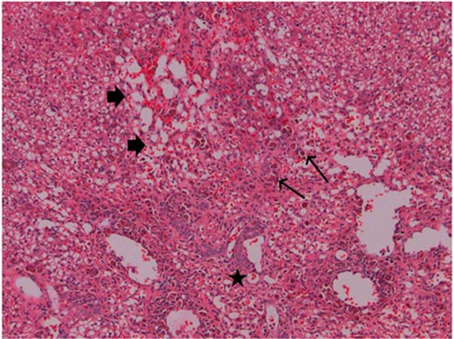
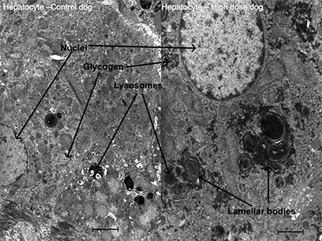
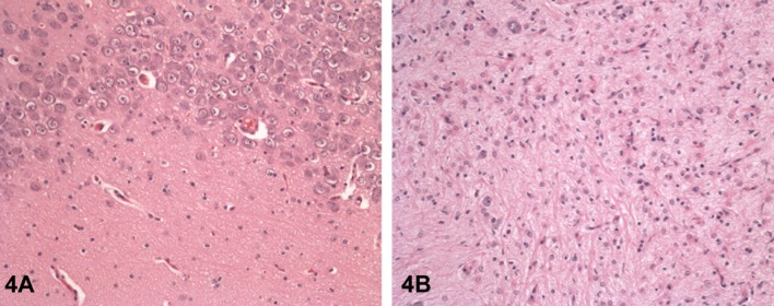
AZD3783, a cationic amphiphilic drug and a potent inhibitor of the 5-hydroxytryptamine (5-HT1B) receptor, was explored as a potential treatment for depression. To support clinical trials, repeat dose toxicity studies in rats and dogs were conducted. Here we report toxicity findings in dogs after dosing from 1 to 3 months. In the 1-month study, there were minimal neuronal vacuolation in the brain, a marked increase in liver enzymes accompanied by hepatocellular degeneration/necrosis and phospholipidosis (PLD), and PLD/cholecystitis in the gallbladder of animals dosed at 47 mg/kg/day. In the 3-month study, neurotoxicity resulted in euthanasia of one animal dosed at 30 mg/kg/day after 86 days. Extensive pathologic changes were seen in all animals in retina epithelium (inclusion bodies), brain (neuronal vacuolation, degeneration, or necrosis and nerve fiber degeneration), spinal ganglia (vacuolation, degeneration, or necrosis), as well as sciatic and optic nerves (degeneration). Pigment-laden macrophages were observed in the lung, kidney, liver, gallbladder, bone marrow, gastrointestinal tract, and lymphoid tissues. Also seen were vitrel and retinal hemorrhage in the eyes. A brain concentration and pathology study showed that the concentration of AZD3783 in the brain was approximately 4 times higher than in the plasma after 4 weeks of dosing, however, they were similar in all regions examined, and did not correlate with areas with pathologic findings. Our findings with AZD3783 in dogs have not been reported previously with other CNS compounds that effect through serotonergic pharmacology.
Keywords: dog; pathology; neurotoxicity; preclinical safety assessment; phospholipidosis; cationic amphiphilic drugs (CADs).
Figures

References
-
- Zhang M, Zhou D, Wang Y, Maier DL, Widzowski DW, Sobotka-Briner CD, Brockel BJ, Potts WM, Shenvi AB, Bernstein PR, and Pierson ME. Preclinical pharmacology and pharmacokinetics of AZD3783, a selective 5-hydroxytryptamine1B receptor antagonist. J Pharmacol Exp Ther. 339: 567–578 2011. - PubMed
-
- Fink KB, and Göthert M. 5-HT receptor regulation of neurotransmitter release. Pharmacol Rev. 59: 360–417 2007. - PubMed
-
- Moret C, and Briley M. The possible role of 5-HT1B/1D receptors in psychiatric disorders and their potential as a target for therapy. Eur J Pharmacol. 404: 1–12 2000. - PubMed
-
- Slassi A. Recent advances in 5-HT1B/1D receptor antagonists and agonists and their potential therapeutic applications. Curr Top Med Chem. 2: 559–574 2002. - PubMed
-
- Varnäs K, Nybert S, Karlsson P, Pierson ME, Kågedal M, Cselényi Z, McCarthy D, Xiao A, Zhang M, Halldin C, and Farde L. Dose-dependent binding of AZD3783 to brain 5- HT1B receptors in non-human primates and human subjects: a positron emission tomography study with [11C]AZ10419369. Psychopharmacology. 213: 533–545 2011. - PubMed
LinkOut - more resources
Full Text Sources
Other Literature Sources