

Magnetoencephalography: Basic principles
- PMID: 24791076
- PMCID: PMC4001219
- DOI: 10.4103/0972-2327.128676
Magnetoencephalography: Basic principles
Abstract
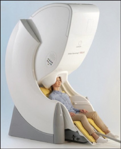
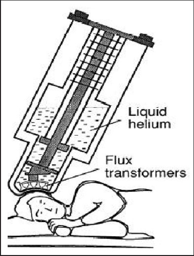
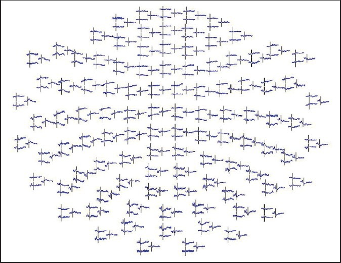
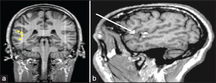
Magnetoencephalography (MEG) is the measurement of the magnetic field generated by the electrical activity of neurons. It is usually combined with a magnetic resonance imaging to get what is called magnetic source imaging. The technology that has helped record these minute magnetic fields is super-conducting quantum interference detector which is like a highly sensitive magnetic field meter. To attenuate the external magnetic noise the MEG is housed inside a magnetically shielded room. The actual sensors recording magnetic fields are magnetometers and/or gradiometers. MEG fields pass through the head without any distortion. This is a significant advantage of MEG over electroencephalography. MEG provides a high spatial and temporal resolution. The recording and identification information should be according to the American Clinical Magnetoencephalography Society guidelines published in 2011. MEG currently has two approved indications in the United States, one is for pre-operative brain mapping and the other is for use in epilepsy surgery. MEG studies have shown functional brain tissue inside brain tumors.
Keywords: Cortical mapping; epilepsy; magnetic source imaging; magnetoencephalography.
Conflict of interest statement
Figures
References
-
- Singh S. Magnetoencephalography. Prog Clin Neurosciences. 2012;26:164–74.
-
- Hämäläinen MS. Basic principles of magnetoencephalography. Acta Radiol Suppl. 1991;377:58–62. - PubMed
-
- Hämäläinen MS. Magnetoencephalography: A tool for functional brain imaging. Brain Topogr. 1992;5:95–102. - PubMed
-
- Hamalainen M, Hari R, Ilmoniemi RJ, Knuutila J, Lounasmaa OV. Magnetoencephalography — Theory, instrumentation, and application to noninvasive studies of the working human brain. Rev Mod Phys. 1993;65:413–97.
-
- Knowlton RC, Elgavish RA, Bartolucci A, Ojha B, Limdi N, Blount J, et al. Functional imaging: II. Prediction of epilepsy surgery outcome. Ann Neurol. 2008;64:35–41. - PubMed
LinkOut - more resources
Full Text Sources
Other Literature Sources
