Review
. 2014 Aug:27:7-13.
doi: 10.1016/j.gde.2014.03.004. Epub 2014 May 8.

Morphomechanics: transforming tubes into organs

Affiliations
Review

Morphomechanics: transforming tubes into organs

Larry A Taber. Curr Opin Genet Dev. 2014 Aug.

Abstract

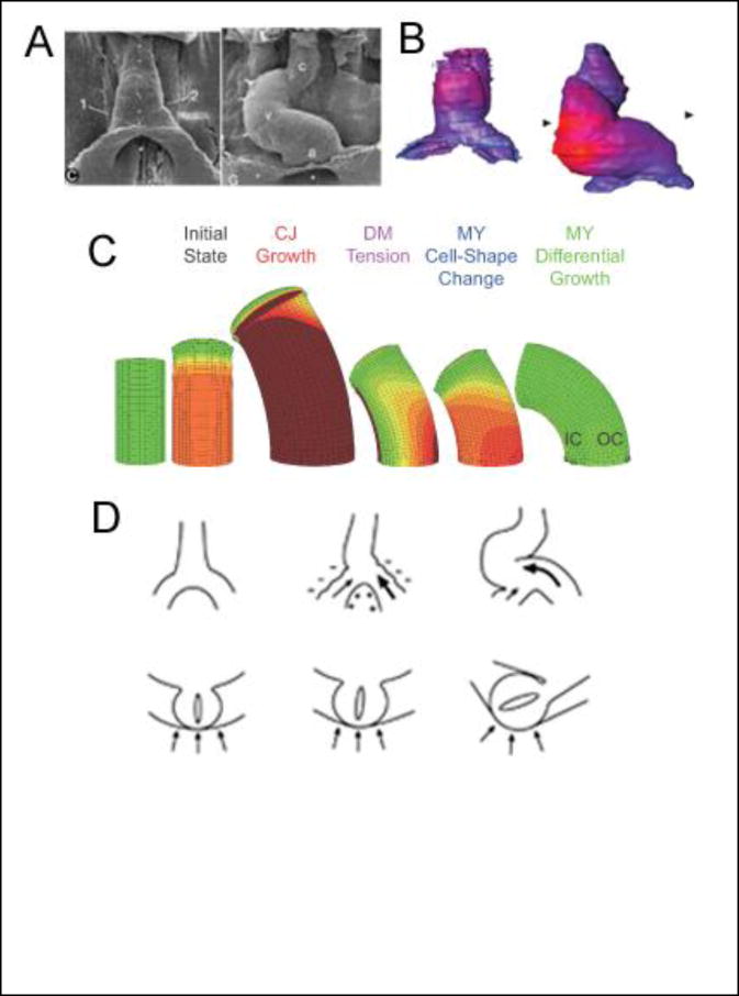
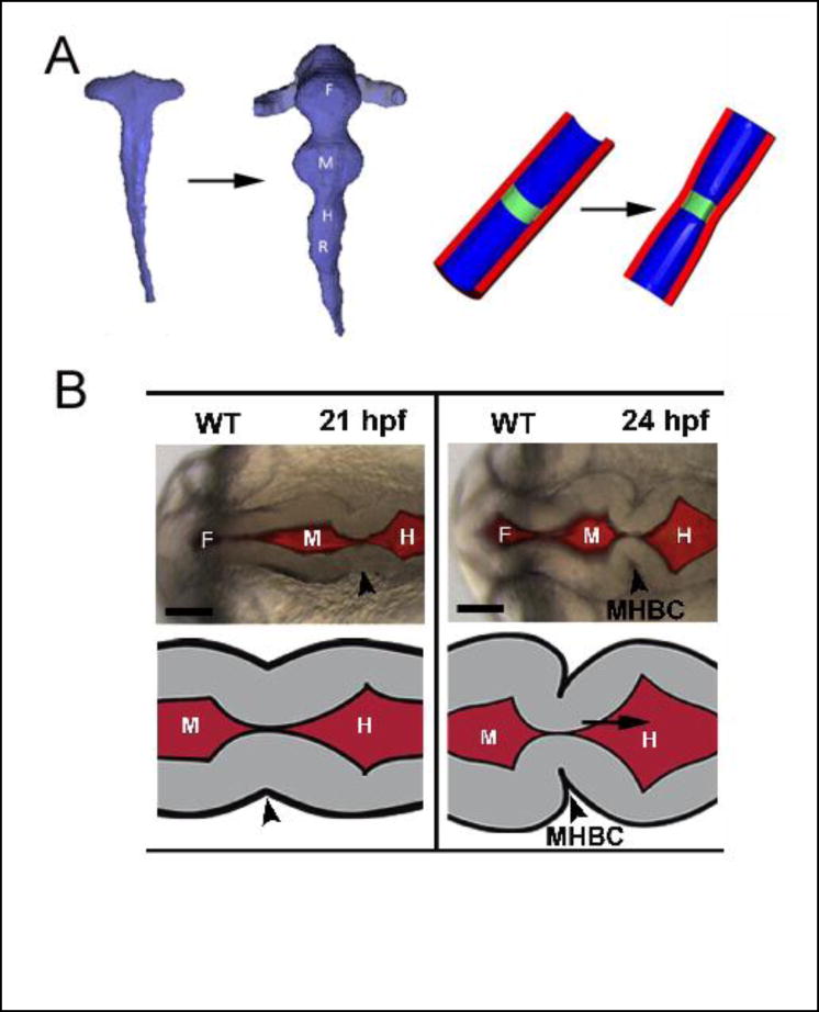
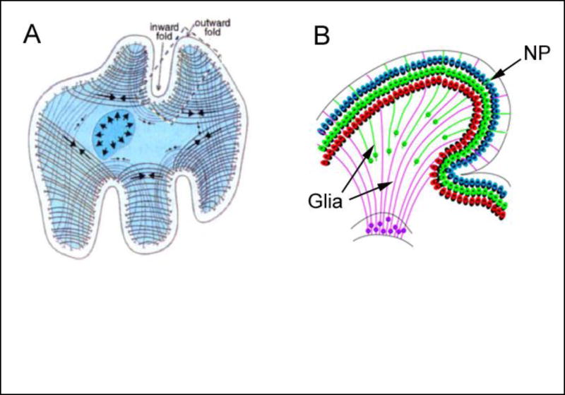
After decades focusing on the molecular and genetic aspects of organogenesis, researchers are showing renewed interest in the physical mechanisms that create organs. This review deals with the mechanical processes involved in constructing the heart and brain, concentrating primarily on cardiac looping, shaping of the primitive brain tube, and folding of the cerebral cortex. Recent studies suggest that differential growth drives large-scale shape changes in all three problems, causing the heart and brain tubes to bend and the cerebral cortex to buckle. Relatively local changes in form involve other mechanisms such as differential contraction. Understanding the mechanics of organogenesis is central to determining the link between genetics and the biophysical creation of form and structure.


Figures

Fig 1
Fig 1. Cardiac looping
(A) Scanning electron micrographs of embryonic chick heart at beginning (left) and end (right) of c-looping (ventral view, stages 10 and 12 of Hamburger and Hamilton [88]). Small dots along the ventral midline of the heart tube move to the outer curvature, illustrating that c-looping consists of ventral bending and rightward torsion. (c = conotruncus; v = ventricle; a = primitive atrium) From [5]. (B) Maps of myocardial cell size at similar stages of development as in (A) (blue = small cells; red = large cells). Dorsal-ventral (inner to outer curvature) gradient in cell size is consistent with a differential growth mechanism for cardiac bending. From [16**]. (C) Computational models for the bending component of c-looping. From the initial state including cardiac jelly (CJ) swelling, the models simulate the following mechanisms: dorsally constrained expansion of the heart tube as CJ continues to grow and swell, dorsal forces exerted on the heart by tension in the rupturing dorsal mesocardium (DM), active cell-shape changes in the myocardium (MY), and differential myocardial growth. Only differential growth yields a bending magnitude consistent with experiments. (IC = inner curvature; OC = outer curvature) From [18**]. (D) Schematic of mechanism for torsional component of c-looping (top row = ventral view; bottom row = cross-sectional view). Left: before looping; center: relatively large force exerted by left omphalomesenteric vein (bold arrow) pushes heart tube slightly rightward; right: compression exerted by splanchnopleure (arrows in cross section) enhances rotation of heart tube. From [22*].
Fig 2
Fig 2. Formation of primary vesicles in brain tube
(A) Chick embryo. Reconstructed brain lumen at stages 10 and 12 (left) and schematic of boundary formation (right). Boundaries between vesicles are created by circumferential actomyosin contraction (green region) at apical side of wall. From [47*]. (B) Zebrafish embryo. Boundary between midbrain and hindbrain (arrowhead) forms in two steps: radial shortening (left) and basal constriction (right) of neuroepithelial cells. (F= forebrain; M = midbrain; H = hindbrain; MHBC = midbrain-hindbrain boundary constriction) From [48*].
Fig 3
Fig 3. Hypotheses for folding of cerebral cortex
(A) Axon tension. Tension generated by axons (arrows on curved lines) draws interconnected regions together, creating gyri (outward folds). From [71**]. (B) Differential growth. Neural progenitors (NP) migrate and spread out long fan-like glial fibers as they enter the cortex, expanding the surface area and creating a gyrus. From [77**].

References

    1. Martin AC. Pulsation and stabilization: contractile forces that underlie morphogenesis. Dev Biol. 2010;341:114–125. - PubMed
    1. Lecuit T, Lenne PF, Munro E. Force generation, transmission, and integration during cell and tissue morphogenesis. Annu Rev Cell Dev Biol. 2011;27:157–184. - PubMed
    1. Nelson CM, Gleghorn JP. Sculpting organs: mechanical regulation of tissue development. Annu Rev Biomed Eng. 2012;14:129–154. - PubMed
    1. Taber LA. Biophysical mechanisms of cardiac looping. International Journal of Developmental Biology. 2006;50:323–332. - PubMed
    1. Manner J. Cardiac looping in the chick embryo: a morphological review with special reference to terminological and biomechanical aspects of the looping process. Anatomical Record. 2000;259:248–262. - PubMed

Publication types

LinkOut - more resources