

doi: 10.1038/leu.2014.152.
Epub 2014 May 5.
Acceleration of Bcr-Abl+ leukemia induced by deletion of JAK2
Affiliations
- PMID: 24791858
- PMCID: PMC4158830
- DOI: 10.1038/leu.2014.152
Item in Clipboard
Acceleration of Bcr-Abl+ leukemia induced by deletion of JAK2
Leukemia.
2014 Sep.
Free PMC article
No abstract available
Figures
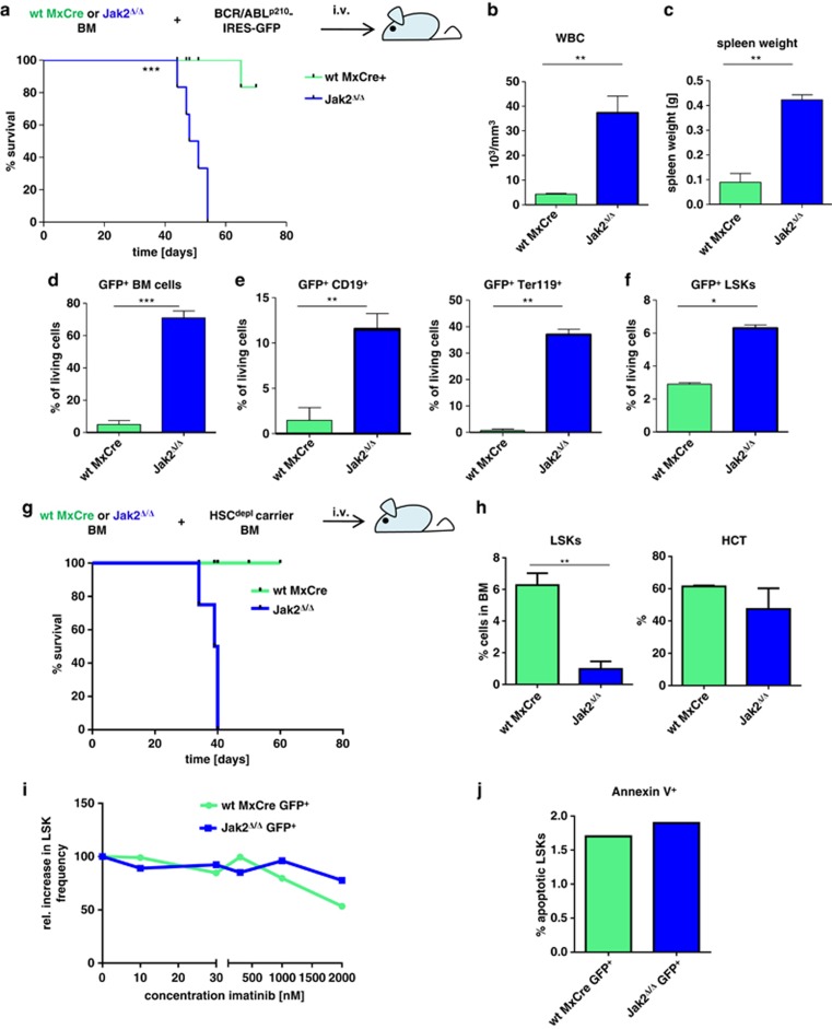
Jak2 deletion accelerates BCR-ABLp210+ leukemia in mice but leads to a reduction of LSKs in normal hematopoiesis. Jak2Δ/Δ and Jak2f/fMxCre+ BM cells (1 × 10⁶) were injected in non-irradiated NSG mice (n=12) (a) Mice that received Jak2Δ/Δ BCR-ABLp210-transformed BM succumb prematurely to leukemia. Short lines indicate individual mice that were killed as control. Mice of the Jak2Δ/Δ cohort display (b) increased peripheral WBCs and (c) spleen weights. (d) Percentages of BCR-ABLp210+/GFP+ cells are increased in BMs of mice that received Jak2Δ/Δ BCR-ABLp210-transformed BM compared with control animals. (e) Jak2Δ/Δ BCR-ABLp210+/GFP+ cells contribute to B-cell and erythroid lineages. (f) Increased percentages of BCR-ABLp210+/GFP+ LSKs in mice that received a Jak2Δ/Δ transplant. (g) Supplementation of Jak2Δ/Δ BM with HSC-depleted carrier BM leads to premature death. Non-transformed Jak2Δ/Δ BM was mixed with high-purity sorted HSC-depleted C57BL/6 J BM cells and injected into lethally irradiated recipients (n=8). Scheme depicts experimental setup. (h) Numbers of LSKs are severely reduced in mice that received a mixture of Jak2Δ/Δ BM and HSC-depleted carrier cells. Hematocrit (HCT) levels remained unaltered upon JAK2 loss. (i) Dose-response curves of BCR-ABLp210+ LSKs incubated for 24 h in the presence of ruxolitinib (300 nM ) and increasing doses of imatinib (ranging from 10 nM to 2 μM ). (j) Frequencies of apoptotic (Annexin V+) LSKs upon imatinib treatment (48 h incubation; 2 μM ). Asterisks denote level of statistical significance as determined by an unpaired t-test: *P⩽0.05; **P⩽0.01; ***P⩽0.001.
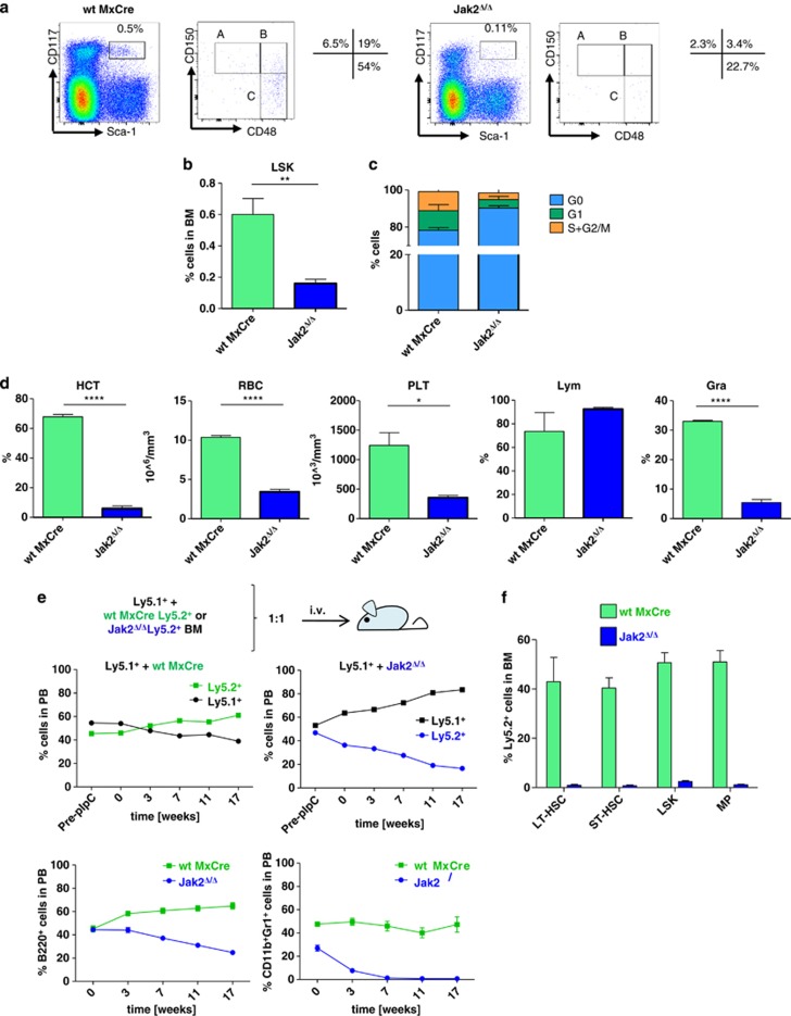
Jak2-deficient BM harbors reduced numbers of HSCs and shows a selective disadvantage in competitive transplantations. Jak2f/fMxCre+ and wtMx1Cre+ mice were treated with poly(I:C) every 3 days for 2 weeks to induce gene deletion (a–c; n=8 for each genotype). (a) Loss of HSCs upon Jak2 deletion. Representative fluorescence-activated cell sorting (FACS) plots indicate reduction of HSC numbers upon Jak2 deletion. After 2 weeks, BM of Jak2Δ/Δ cells displayed significantly reduced numbers of LSK and fraction A, B and C cells. Upper panels: Representative FACS plot depicting LSK cells that were further subdivided by CD150 and CD48 expression. Percentages of cells belonging to fraction A, B or C are provided next to the plot. (b) Summary of LSK numbers in Jak2Δ/Δ and wtMx1Cre+ mice. BM cells were analyzed as described in (a). (c) Higher numbers of HSCs in G0 phase in Jak2Δ/Δ mice. BM cells of Jak2Δ/Δ and wtMx1Cre+ mice were analyzed for Ki67 and 4′,6-diamidino-2-phenylindole incorporation. The majority of Jak2Δ/Δ cells underwent the G0 phase of the cell cycle. (d) Analyses of peripheral blood (PB) of poly(I:C)-treated wtMx1Cre+ and Jak2Δ/Δ mice. Hematocrit (HCT), RBCs and PLT counts as well as percentages of lymphocytes and granulocytes are summarized in bar graphs. (e) BM of Ly5.1+ mice was mixed with either Jak2f/fMxCre+ or wtMx1Cre (both Ly5.2+) BM cells and injected intravenously into lethally irradiated Ly5.1+ mice. Post transplantation, mice received poly (I:C) to delete Jak2. Upper panels: percentages of peripheral blood cells expressing Ly5.1 or Ly5.2 of mice that received a Ly5.1/wtMx1Cre (left panel) or a Ly5.1/Jak2f/fMxCre+ (right panel) mixture of cells (n=9 each). Lower panels: percentages of CD11b+Gr-1+Ly.5.2+ (left panel) and B220+Ly5.2+ (right panel) cells in the peripheral blood of mice that received a Ly5.1/wt or a Ly5.1/Jak2f/fMxCre+ mixture of cells are shown. (f) Numbers of Jak2Δ/Δ HSCs and MPs are markedly reduced in a competitive setting with wt cells. Asterisks denote level of statistical significance as determined by an unpaired t-test: *P⩽0.05; **P⩽0.01; ****P⩽0.0001.
Similar articles
-
Janus kinase 2 regulates Bcr-Abl signaling in chronic myeloid leukemia.Leukemia. 2011 Mar;25(3):463-72. doi: 10.1038/leu.2010.287. Epub 2010 Dec 24. Leukemia. 2011. PMID: 21183952 Free PMC article.
-
BCR-ABL uncouples canonical JAK2-STAT5 signaling in chronic myeloid leukemia.Nat Chem Biol. 2012 Jan 29;8(3):285-93. doi: 10.1038/nchembio.775. Nat Chem Biol. 2012. PMID: 22286129
-
BCR-ABL but not JAK2 V617F inhibits erythropoiesis through the Ras signal by inducing p21CIP1/WAF1.J Biol Chem. 2010 Oct 8;285(41):31774-82. doi: 10.1074/jbc.M110.118653. Epub 2010 Jul 27. J Biol Chem. 2010. PMID: 20663870 Free PMC article.
-
Concurrent JAK2-Positive Myeloproliferative Disorder and Chronic Myelogenous Leukemia: A Novel Entity? A Case Report With Review of the Literature.J Investig Med High Impact Case Rep. 2019 Jan-Dec;7:2324709619832322. doi: 10.1177/2324709619832322. J Investig Med High Impact Case Rep. 2019. PMID: 30803277 Free PMC article. Review.
-
Hematopoietic stem cells in chronic myeloid leukemia.Arch Med Res. 2003 Nov-Dec;34(6):496-506. doi: 10.1016/j.arcmed.2003.09.005. Arch Med Res. 2003. PMID: 14734089 Review.
Cited by
-
The KRAS-G12D mutation induces metabolic vulnerability in B-cell acute lymphoblastic leukemia.iScience. 2022 Feb 7;25(3):103881. doi: 10.1016/j.isci.2022.103881. eCollection 2022 Mar 18. iScience. 2022. PMID: 35243242 Free PMC article.
-
Inhibition of STAT5: a therapeutic option in BCR-ABL1-driven leukemia.Oncotarget. 2014 Oct 30;5(20):9564-76. doi: 10.18632/oncotarget.2465. Oncotarget. 2014. PMID: 25333255 Free PMC article. Review.
-
JAK2/STAT5 inhibition by nilotinib with ruxolitinib contributes to the elimination of CML CD34+ cells in vitro and in vivo.Blood. 2014 Aug 28;124(9):1492-501. doi: 10.1182/blood-2013-12-545640. Epub 2014 Jun 23. Blood. 2014. PMID: 24957147 Free PMC article.
-
Decreased NK-cell tumour immunosurveillance consequent to JAK inhibition enhances metastasis in breast cancer models.Nat Commun. 2016 Jul 13;7:12258. doi: 10.1038/ncomms12258. Nat Commun. 2016. PMID: 27406745 Free PMC article.
-
Carboplatin-Induced Thrombocytopenia through JAK2 Downregulation, S-Phase Cell Cycle Arrest and Apoptosis in Megakaryocytes.Int J Mol Sci. 2022 Jun 3;23(11):6290. doi: 10.3390/ijms23116290. Int J Mol Sci. 2022. PMID: 35682967 Free PMC article.
References
-
- Harrison C, Kiladjian J-J, Al-Ali HK, Gisslinger H, Waltzman R, Stalbovskaya V, et al. JAK inhibition with ruxolitinib versus best available therapy for myelofibrosis. N Engl J Med. 2012;366:787–798. - PubMed
-
- Harry BL, Eckhardt SG, Jimeno A. JAK2 inhibition for the treatment of hematologic and solid malignancies. Expert Opin Investig Drugs. 2012;21:637–655. - PubMed
Publication types
MeSH terms
Substances
Grants and funding
LinkOut - more resources
Full Text Sources
Other Literature Sources
Medical
Molecular Biology Databases
Miscellaneous
