A single injection of intravitreal ranibizumab in the treatment of choroidal neovascularisation secondary to optic nerve head drusen in a child
- PMID: 24792030
- PMCID: PMC4025440
- DOI: 10.1136/bcr-2014-204456
A single injection of intravitreal ranibizumab in the treatment of choroidal neovascularisation secondary to optic nerve head drusen in a child
Abstract
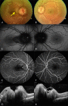
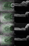
Optic nerve head drusen are acellular, calcified deposits which may be found in buried or exposed drusen form. Choroidal neovascularisation secondary to optic nerve head drusen is rarely seen in childhood. This case report summarises the clinical and therapeutic outcomes of a 13-year-old girl with unilateral choroidal neovascularisation secondary to optic nerve head drusen. The patient was successfully treated with a single intravitreal ranibizumab injection. After a month from the injection the visual acuity increased dramatically and maintained at the same level during 9 months of follow-up time. There was no complication related to the injection.
Figures


References
-
- Rodriguez PF, Gili P, Rios MDM. Ophthalmic features of optic disc drusen. Ophthalmologica 2012;228:59–66 - PubMed
-
- Auw-Haedrich C, Staubach F, Witschel H. Optic disk drusen. Surv Ophthalmol 2002;47:515–32 - PubMed
-
- Delyfer M, Rougier M, Fourmaux E, et al. Laser photocoagulation for choroidal neovascular membrane associated with optic disc drusen. Acta Ophthalmol Scand 2004;82:236–8 - PubMed
-
- Adan A, Corcóstegui B. Surgical removal of peripapillary choroidal neovascularization associated with optic nerve drusen. Retina 2004;24:739–45 - PubMed
-
- Torrent R, Loureiro R, Travassos A, et al. Bilateral CNV associated with optic nerve drusen treated with photodynamic therapy with verteporfin. Eur J Ophthalmol 2004;14:434–7 - PubMed
Publication types
MeSH terms
Substances
LinkOut - more resources
Full Text Sources
Other Literature Sources
