

Mammalian fetal cardiac regeneration after myocardial infarction is associated with differential gene expression compared with the adult
- PMID: 24792251
- PMCID: PMC4010943
- DOI: 10.1016/j.athoracsur.2014.01.013
Mammalian fetal cardiac regeneration after myocardial infarction is associated with differential gene expression compared with the adult
Abstract
Background: In adults, myocardial infarction (MI) results in a brisk inflammatory response, myocardium loss, and scar formation. We have recently reported the first mammalian large-animal model of cardiac regeneration after MI in fetal sheep. We hypothesize that the ability of the fetus to regenerate functional myocardium after MI is owing to differential gene expression regulating the response to MI in the fetus compared with the adult.
Methods: Myocardial infarction was created in adult (n=4) or early gestation fetal (n=4) sheep. Tissue was harvested after 3 or 30 days, and RNA was extracted for microarray, followed by principal component analysis and global gene expression analysis for the following gene ontology terms: response to wounding, inflammatory response, extracellular matrix, cell cycle, cell migration, cell proliferation, and apoptosis.
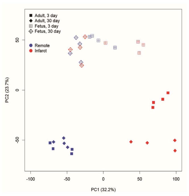
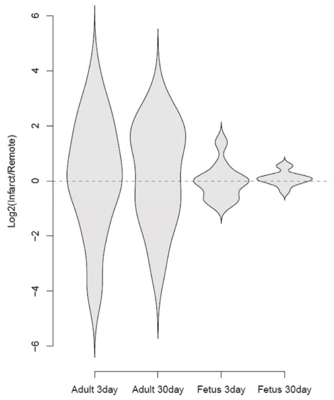
Results: Principal component analysis demonstrated that the global gene expression pattern in adult infarcts was distinctly different from the uninfarcted region at 3 days and remained different at 30 days after MI. In contrast, gene expression in the fetal infarct was different from the uninfarcted region at 3 days, but by 30 days it returned to a baseline expression pattern similar to the uninfarcted region. Three days after MI there was an increase in the expression of genes related to all gene ontology terms in fetal and adult infarcts, but this increase was much more pronounced in adults. By 30 days, the fetal gene expression returned to baseline, whereas in the adult it remained significantly elevated.
Conclusions: These data demonstrate that the global gene expression pattern is dramatically different in the fetal regenerative response to MI compared with the adult response and may partly be responsible for the regeneration.
Copyright © 2014 The Society of Thoracic Surgeons. Published by Elsevier Inc. All rights reserved.
Figures


Comment in
-
Invited commentary.Ann Thorac Surg. 2014 May;97(5):1650-1. doi: 10.1016/j.athoracsur.2014.01.024. Ann Thorac Surg. 2014. PMID: 24792252 No abstract available.
References
-
- Go AS, Mozaffarian D, Roger VL, Benjamin EJ, Berry JD, Borden WB, et al. Executive summary: heart disease and stroke statistics--2013 update: a report from the American Heart Association. Circulation. 2013;127:143–52. - PubMed
-
- Bolognese L, Ducci K, Angioli P, Falsini G, Liistro F, Baldassarre S, et al. Elevations in troponin I after percutaneous coronary interventions are associated with abnormal tissue-level perfusion in high-risk patients with non-ST-segment-elevation acute coronary syndromes. Circulation. 2004;110:1592–7. - PubMed
-
- Gheorghiade M, Bonow RO. Chronic heart failure in the United States: a manifestation of coronary artery disease. Circulation. 1998;97:282–9. - PubMed
-
- Levy D, Kenchaiah S, Larson MG, Benjamin EJ, Kupka MJ, Ho KK, et al. Long-term trends in the incidence of and survival with heart failure. The New England journal of medicine. 2002;347:1397–402. - PubMed
-
- Sutton MG, Sharpe N. Left ventricular remodeling after myocardial infarction: pathophysiology and therapy. Circulation. 2000;101:2981–8. - PubMed
Publication types
MeSH terms
Substances
Grants and funding
LinkOut - more resources
Full Text Sources
Other Literature Sources
Medical
