. 2014 Jun;5(3):365-74.
doi: 10.1007/s13244-014-0329-1. Epub 2014 May 3.

Radiologic manifestations of angioedema

Affiliations

Radiologic manifestations of angioedema

Kousei Ishigami et al. Insights Imaging. 2014 Jun.

Abstract

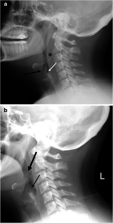
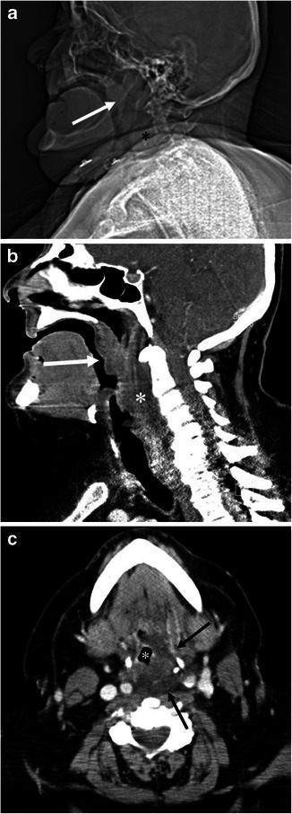
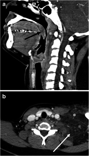
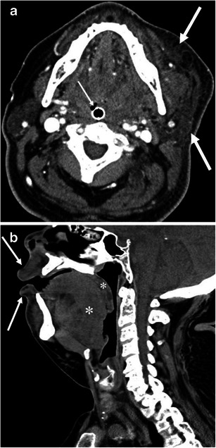
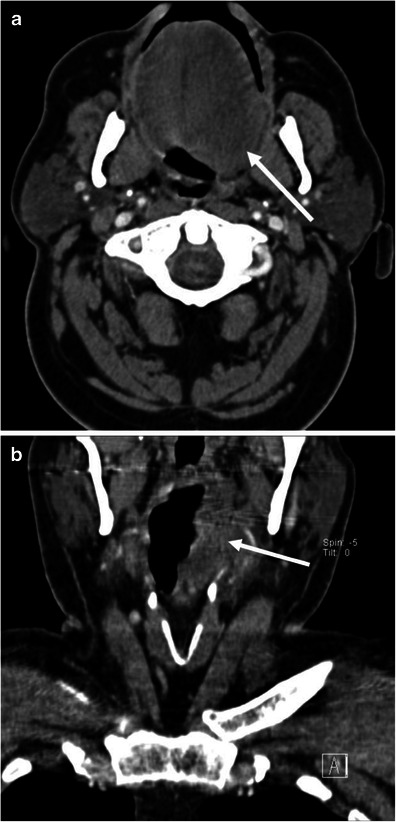
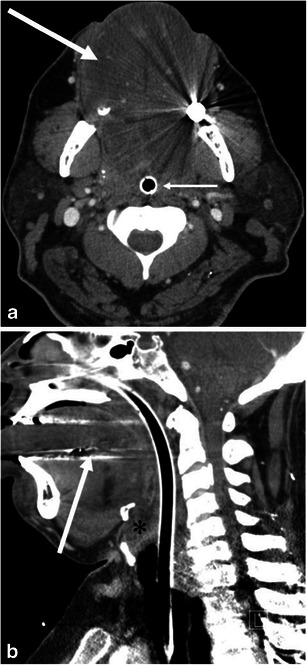
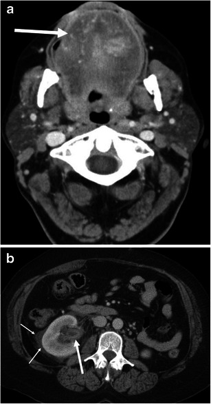
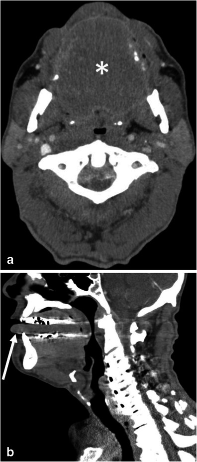
Objectives: The purpose of this pictorial review is to present imaging findings of angioedema involving the various organs.

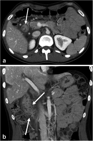
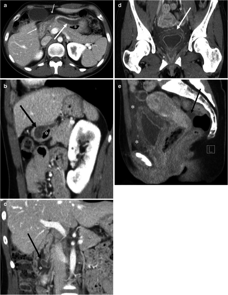
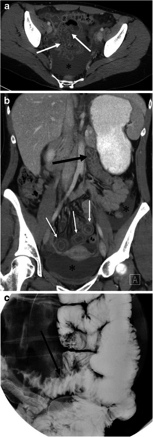
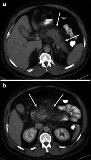
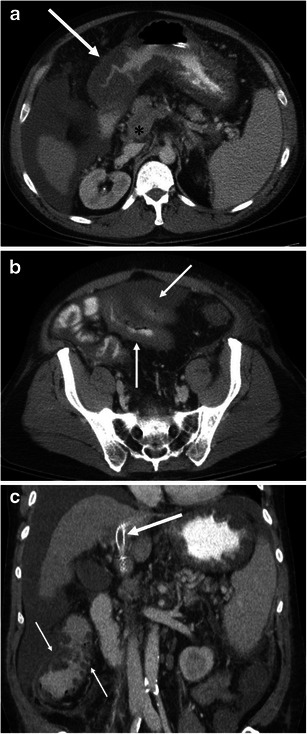
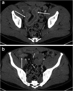
Conclusion: The role of imaging for patients with angioedema includes the evaluation of the upper airway for obstruction and the exclusion of other possible aetiologies, such as neoplastic or infectious processes. Glossomegaly is a common finding of head and neck angioedema. Angioedema may involve organ systems beyond the superficial regions and the head and neck including the gastrointestinal and genitourinary tracts. Angioedema of the visceral organs is often accompanied by adjacent fluid, and it is commonly diffuse or concentric but can also be multifocal and asymmetric.

Teaching points: • The evaluation of the upper airway obstruction is important for head and neck angioedema. • Glossomegaly with decreased attenuation is common in head and neck angioedema. • Angioedema of the visceral organs can be multifocal and asymmetric. • Angioedema of the visceral organs is often accompanied by adjacent fluid. • It is important to include clinical and laboratory findings for the diagnosis of angioedema.


Figures

Fig. 1
Fig. 1
A 33-year-old female with hereditary angioedema (HAE). Soft tissue swelling of the neck during recurrent angioedema attacks. a The patient complained of neck swelling. Lateral radiograph of the neck shows thickening of the retropharyngeal soft tissue (asterisk), enlargement of the epiglottis with narrowing of the vallecula (large arrow), and thickening of the aryepiglottic folds (small arrow). b Six months later, the patient again complained of neck swelling and swallowing difficulty. Lateral view of the neck plain film shows enlargement of the epiglottis (large arrow, thumb sign) and thickening of the aryepiglottic fold (small arrow). No retropharyngeal soft tissue thickening is noted
Fig. 2
Fig. 2
A 68-year-old male with angiotensin converting enzyme (ACE) inhibitor-induced angioedema involving the soft palate, pharynx and larynx. a Scout view of the neck computed tomography (CT) shows enlargement of the soft palate (arrow) and thickening of the retropharyngeal soft tissue (asterisk). b The sagittal reformatted contrast enhanced (CE)-CT shows marked swelling of the soft palate (arrow) and posterior pharyngeal wall (asterisk) with decreased attenuation. c The axial CE-CT image shows swelling of the left posterolateral retropharyngeal space (arrows), and narrow displaced pharyngeal airway (asterisk)
Fig. 3
Fig. 3
A 24-year-old female with systemic lupus erythaematosus and anti-phospholipid syndrome presenting with acquired angioedema. a The sagittal reformatted CE-CT shows diffuse retropharyngeal (asterisk) and soft palate oedema (arrow) causing upper airway encroachment at the level of the oropharynx. b The axial CE-CT shows extensive soft tissue oedema with fat stranding of the left lower neck (arrow)
Fig. 4
Fig. 4
A 64-year-old male with ACE inhibitor-induced angioedema with airway obstruction. a The axial CE-CT image shows left hemifacial swelling with subcutaneous fat stranding (large arrows). The patient is status post placement of an endotracheal tube (small arrow). b One year later, the patient developed recurrent angioedema after tooth extraction. The sagittal reformatted image of the unenhanced CT shows marked swelling of the tongue base and soft palate (asterisks). Also noted is swelling of the lips (arrows)
Fig. 5
Fig. 5
A 62-year-old male with ACE inhibitor-induced angioedema. a The axial image of CE-CT demonstrates asymmetric left-sided tongue oedema (arrow). b The coronal reformatted image of CE-CT shows extension of oedema into the left pharyngeal wall (arrow)
Fig. 6
Fig. 6
A 49-year-old male with ACE inhibitor-induced angioedema. a The axial CE-CT shows diffuse enlargement of the tongue with decreased attenuation due to oedema (large arrow). Small arrow indicates the endotracheal tube. b Sagittal CE-CT shows glossomegaly (arrow) and marked laryngeal oedema (asterisk). The endotracheal tube is inserted by the trans-nasal approach because of marked glossomegaly
Fig. 7
Fig. 7
A 54-year-old female with ACE inhibitor-induced angioedema. a The axial CE-CT of the neck shows diffuse enlargement of the tongue (arrow). Vessels are recognised within the tongue, reflecting decreased attenuation due to oedema. b The axial CE-CT of the abdomen on the same date shows thickened and contrast-enhancing uroepithelium of the renal pelvis (large arrow). Perinephric fluid is also noted (small arrows). No stone disease was recognised in the ureter or urinary bladder (not shown). Urinalysis was unremarkable except for microscopic haematuria, which returned to normal 3 days later. Follow-up CT 1 month later showed complete resolution of renal pelvic wall thickening and perinephric fluid (not shown)
Fig. 8
Fig. 8
A 64-year-old male with amyloidosis presenting with diffuse tongue swelling. a The axial CE-CT shows the tongue to be diffusely swollen (asterisk). Note the attenuation of the tongue is soft tissue density. Glossomegaly caused by angioedema typically shows low density (see Figs. 6 and 7). b The sagittal CE-CT shows diffuse tongue swelling with anterior protrusion of the tip of the tongue (arrow)
Fig. 9
Fig. 9
A 26-year-old female with HAE involving the 2nd portion of the duodenum. a The axial CE-CT shows the duodenal wall to be thickened (arrow) with surrounding free fluid (asterisks). b The coronal reformatted image shows oedematous thickening of the duodenal folds (arrows)
Fig. 10
Fig. 10
A 27-year-old female with HAE involving the stomach, ampulla of Vater, urinary bladder, and small and large bowel (not shown). a The axial CE-CT shows marked submucosal oedema of the anterior wall of the stomach (arrow). Asterisk indicates the lumen of the stomach. Fluid is noted adjacent to the stomach and duodenum (small arrows). b The sagittal reformatted image clearly shows asymmetrical gastric wall involvement of angioedema (arrow). Asterisk indicates the lumen of the stomach. c Coronal reformatted image shows marked oedematous swelling of the ampulla of Vater (arrow). Asterisk indicates the duodenal lumen. d The coronal reformatted image of the pelvis shows mucosal enhancement and extensive submucosal oedema of the urinary bladder (arrow). Urinalysis was unremarkable without evidence of urinary tract infection. e The sagittal reformatted image shows fluid around the urinary bladder in the extraperitoneal space (asterisks). Arrow indicates a small amount of intraperitoneal free fluid in the cul-de-sac
Fig. 11
Fig. 11
A 29-year-old female with ACE inhibitor-induced angioedema involving multiple small bowel loops. a The axial CE-CT shows oedematous thickening of the small bowel folds with mucosal enhancement (arrows). A moderate amount of intraperitoneal free fluid is noted in the pelvis (asterisk). b The coronal reformatted image demonstrates oedematous wall thickening of the proximal jejunum and intrapelvic small bowel. The proximal jejunum (large arrow) shows oedematous thickening of the jejunal folds. The short axis section of the involved intrapelvic small bowel shows the halo sign, representing a low-density submucosal layer with mucosal and subserosal enhancement (small arrows). Asterisks denote intraperitoneal free fluid. c Spot film of single-contrast small bowel barium study shows straight small bowel folds thickening (arrow) and a stack of coins appearance
Fig. 12
Fig. 12
A 44-year-old male with HAE presenting with acute pancreatitis. a The axial CE-CT image shows fat stranding around the pancreatic body and tail (arrows). b At the level of the pancreatic head, there is more extensive peripancreatic fat stranding (arrows). Wall thickening of the 2nd portion of the duodenum (asterisk) is seen, although it could be caused by pancreatitis
Fig. 13
Fig. 13
A 55-year-old male with liver cirrhosis presenting with portal venous thrombus and resultant small bowel ischaemia and congestion of the gastrointestinal tracts. a The venous phase of the axial CE-CT shows marked oedematous wall thickening of the gastric antrum (arrow). The splenoportal confluence (asterisk) is poorly opacified. Ascites and splenomegaly are also noted. b The axial CE-CT at the level of the pelvis shows wall thickening of the small bowel (arrows). Laparotomy found necrosis of the small bowel. c The coronal reformatted image shows an occluded transjugular intrahepatic portosystemic shunt (TIPS) stent (large arrow). The portal vein is not opacified at the level of the hepatic hilum due to thrombus. The ascending colon showed oedematous wall thickening (small arrows)
Fig. 14
Fig. 14
A 42-year-old male with Crohn’s disease. a The axial CE-CT shows wall thickening and hyperenhancement of the terminal ileum (arrows). The layer of wall enhancement is thick and irregular. b The terminal ileum near the ileocecal valve shows wall enhancement and luminal narrowing (arrow). Creeping fat (asterisk) and engorgement of the vasa recta (small arrows) are noted

References

    1. Quinke HE. Über akutes umschriebenes Hautödem. Monatsh Prakt Dermatol. 1882;1:129–131.
    1. de Graaff LC, van Essen M, Schipper EM, Boom H, Duschek EJ. Unnecessary surgery for acute abdomen secondary to angiotensin-converting enzyme inhibitor use. Am J Emerg Med. 2012;30(8):1607–1612. doi: 10.1016/j.ajem.2011.10.028. - DOI - PubMed
    1. Frigas E, Nzeako UC. Angioedema. Pathogenesis, differential diagnosis, and treatment. Clin Rev Allergy Immunol. 2002;23(2):217–231. doi: 10.1385/CRIAI:23:2:217. - DOI - PubMed
    1. Vleeming W, van Amsterdam JG, Stricker BH, de Wildt DJ. ACE inhibitor-induced angioedema. Incidence, prevention and management. Drug Saf. 1998;18(3):171–188. doi: 10.2165/00002018-199818030-00003. - DOI - PubMed
    1. Bork K, Meng G, Staubach P, Hardt J. Hereditary angioedema: new findings concerning symptoms, affected organs, and course. Am J Med. 2006;119(3):267–274. doi: 10.1016/j.amjmed.2005.09.064. - DOI - PubMed

LinkOut - more resources