. 2014 Jun 25;171(1-2):102-11.
doi: 10.1016/j.vetmic.2014.04.005. Epub 2014 Apr 13.

Feline fecal virome reveals novel and prevalent enteric viruses

Affiliations

Feline fecal virome reveals novel and prevalent enteric viruses

Terry Fei Fan Ng et al. Vet Microbiol. .

Abstract

Humans keep more than 80 million cats worldwide, ensuring frequent exposure to their viruses. Despite such interactions the enteric virome of cats remains poorly understood. We analyzed a fecal sample from a single healthy cat from Portugal using viral metagenomics and detected five eukaryotic viral genomes. These viruses included a novel picornavirus (proposed genus "Sakobuvirus") and bocavirus (feline bocavirus 2), a variant of feline astrovirus 2 and sequence fragments of a highly divergent feline rotavirus and picobirnavirus. Feline sakobuvirus A represents the prototype species of a proposed new genus in the Picornaviridae family, distantly related to human salivirus and kobuvirus. Feline astroviruses (mamastrovirus 2) are the closest known relatives of the classic human astroviruses (mamastrovirus 1), suggestive of past cross-species transmission. Presence of these viruses by PCR among Portuguese cats was detected in 13% (rotavirus), 7% (astrovirus), 6% (bocavirus), 4% (sakobuvirus), and 4% (picobirnavirus) of 55 feline fecal samples. Co-infections were frequent with 40% (4/10) of infected cats shedding more than one of these five viruses. Our study provides an initial description of the feline fecal virome indicating a high level of asymptomatic infections. Availability of the genome sequences of these viruses will facilitate future tropism and feline disease association studies.

Keywords: Enteric virus; Felis catus; Metagenomics; Sakobuvirus; Virome.


Figures

Fig. 1
Fig. 1
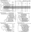
Phylogenetic and genomic analysis of five feline enteric viruses. The Bayesian inference trees were conducted based on A) 3D RdRp protein depicting relationships among representative members of the family Picornaviridae, a table detailing genomic features of the reference genomes based on previously published literature (Lau et al., 2012b; Sweeney et al., 2012). Symbols for protease were noted as follows: Y, presence of a protease motif in L; Y/N, presence of L but L is not protease; N, absence of L. Other motifs are described in the main text. B) the complete ORF2 capsid protein of representative astroviruses C) the complete non-structural protein (NS1) of bocaviruses, D) the partial intermediate capsid protein of rotavirus Segment 6, and E) the partial RdRp protein of picobirnavirus Segment 2. Posterior probabilities of the Bayesian analysis (>95%) are shown next to each node. Scale bar indicates amino acid substitutions per site.
Fig. 2
Fig. 2
Genome organization, identity plot and pairwise sequence comparison analyses of three feline virus genomes: A) feline sakobuvirus A, B) feline astrovirus Viseu, and C) feline bocavirus 2. Cleavage site predictions of the sakobuvirus are shown below its genome. For each virus, three identity plots are shown below the genome organization, comparing the coding regions for each virus with three related viruses based on phylogenetic analyses in Fig. 1. The identity values were color-coded: green, 100%; light green, 30-100%, red, <30%.
Fig. 3
Fig. 3
Predicted RNA secondary structure of the 5′ and 3′ UTR of the feline sakobuvirus A (FeAstV-A) and the 3′ UTR of the feline astrovirus Viseu. For FeAstV-A 5′ UTR, the type IV IRES has been annotated according to picornavirus conventions. Domains are labeled II and III; individual helical segments are labeled II1, II2, III1, and III2, etc.; and individual hairpins are labeled IIIa and IIIb, etc. to maintain the continuity of the current nomenclature. The positions of conserved domains and the polyprotein AUG start codon are indicated by shaded boxes. For FeAstV-A 3′ UTR, detailed structure of the 'barbell-like' formation between nucleotide 7712 and 7775 was shown. Grey boxes indicate identical nucleotides in members of genera Avihepatovirus, Kobuvirus, “Gallivirus” and “Passerivirus”. The presence of poly(Y) tract characteristic of this barbell-like structure is indicated.
Fig. 4
Fig. 4
Venn diagram analysis of the five enteric viruses in fecal samples from 55 cats. Co-infections of more than one virus were annotated.

References

    1. Adams MJ, King AM, Carstens EB. Ratification vote on taxonomic proposals to the International Committee on Taxonomy of Viruses (2013) Archives of virology. 2013;158:2023–2030. - PubMed
    1. Allander T, Tammi MT, Eriksson M, Bjerkner A, Tiveljung-Lindell A, Andersson B. Cloning of a human parvovirus by molecular screening of respiratory tract samples. Proceedings of the National Academy of Sciences of the United States of America. 2005;102:12891–12896. - PMC - PubMed
    1. Allison AB, Kohler DJ, Fox KA, Brown JD, Gerhold RW, Shearn-Bochsler VI, Dubovi EJ, Parrish CR, Holmes EC. Frequent cross-species transmission of parvoviruses among diverse carnivore hosts. Journal of virology. 2013;87:2342–2347. - PMC - PubMed
    1. Ayukekbong J, Lindh M, Nenonen N, Tah F, Nkuo-Akenji T, Bergström T. Enteric viruses in healthy children in cameroon: Viral load and genotyping of norovirus strains. Journal of medical virology. 2011;83:2135–2142. - PubMed
    1. Chung JY, Kim SH, Kim YH, Lee MH, Lee KK, Oem JK. Detection and genetic characterization of feline kobuviruses. Virus genes 2013 - PMC - PubMed

Publication types

Associated data