. 2014 Jul 1;307(1):L38-47.
doi: 10.1152/ajplung.00318.2013. Epub 2014 May 2.

Human airway smooth muscle maintain in situ cell orientation and phenotype when cultured on aligned electrospun scaffolds

Affiliations

Human airway smooth muscle maintain in situ cell orientation and phenotype when cultured on aligned electrospun scaffolds

G E Morris et al. Am J Physiol Lung Cell Mol Physiol. .

Erratum in

Abstract

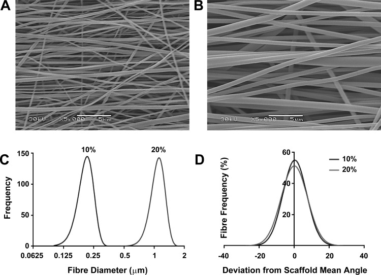
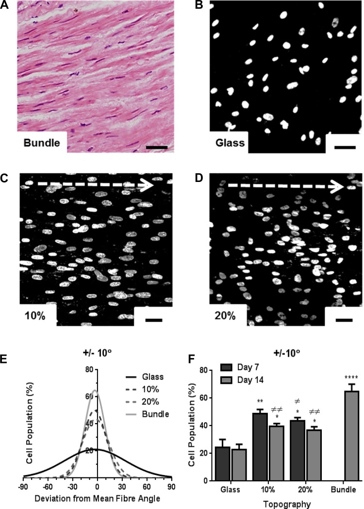
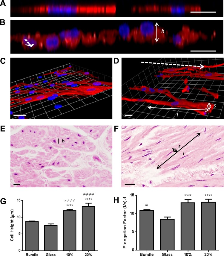
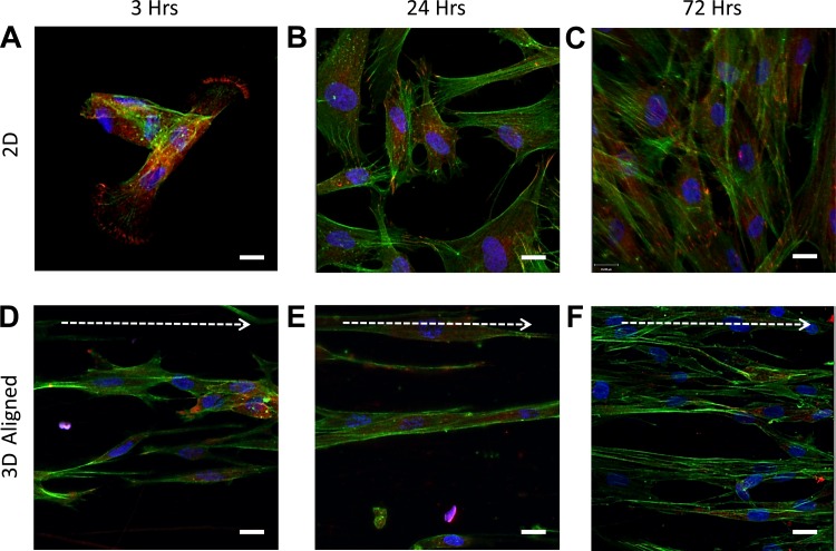
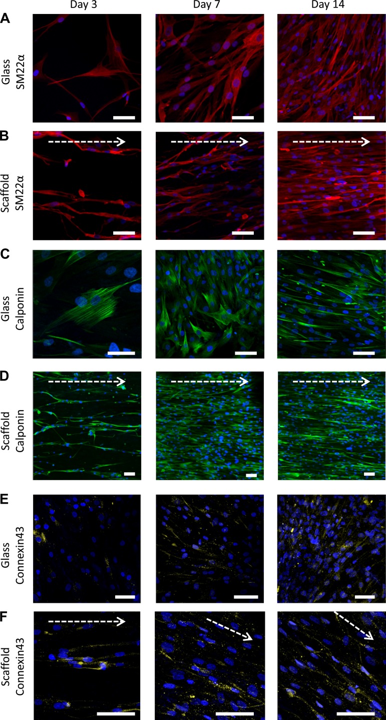
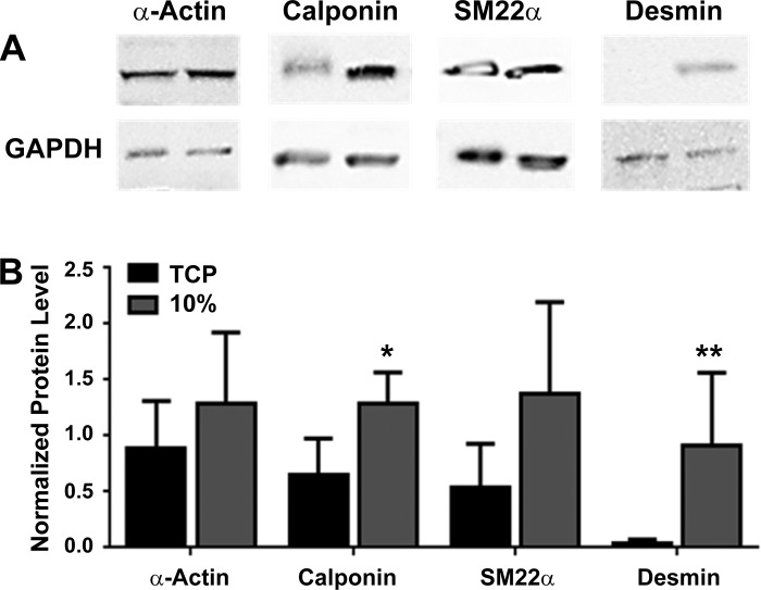
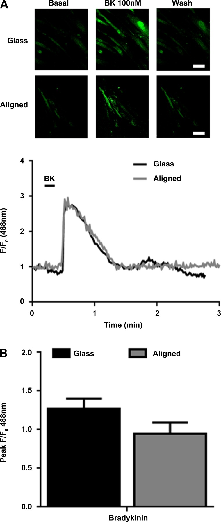
Human airway smooth muscle (HASM) contraction plays a central role in regulating airway resistance in both healthy and asthmatic bronchioles. In vitro studies that investigate the intricate mechanisms that regulate this contractile process are predominantly conducted on tissue culture plastic, a rigid, 2D geometry, unlike the 3D microenvironment smooth muscle cells are exposed to in situ. It is increasingly apparent that cellular characteristics and responses are altered between cells cultured on 2D substrates compared with 3D topographies. Electrospinning is an attractive method to produce 3D topographies for cell culturing as the fibers produced have dimensions within the nanometer range, similar to cells' natural environment. We have developed an electrospun scaffold using the nondegradable, nontoxic, polymer polyethylene terephthalate (PET) composed of uniaxially orientated nanofibers and have evaluated this topography's effect on HASM cell adhesion, alignment, and morphology. The fibers orientation provided contact guidance enabling the formation of fully aligned sheets of smooth muscle. Moreover, smooth muscle cells cultured on the scaffold present an elongated cell phenotype with altered contractile protein levels and distribution. HASM cells cultured on this scaffold responded to the bronchoconstrictor bradykinin. The platform presented provides a novel in vitro model that promotes airway smooth muscle cell development toward a more in vivo-like phenotype while providing topological cues to ensure full cell alignment.

Keywords: airway smooth muscle; aligned fibers; electrospinning; in vitro model; tissue engineering.


Figures

Fig. 1.
Fig. 1.
Polyethylene terephthalate (PET) electrospun scaffolds can be manipulated to form aligned nano- and microfiber scaffolds. PET [either 10 or 20% (wt/vol)] was electrospun onto a rotating mandrel (600 m/min) to produce aligned fibers with diameters in the nano- or micrometer range. Representative scanning electron microscope images of the 10% (nano) and 20% (micro) scaffolds are shown in A and B, respectively. Intrascaffold fiber diameter distributions are shown in C (n = 480 from 3 separate scaffolds), and intrascaffold fiber alignments are shown in D (deviation of individual fiber angle from scaffold's mean fiber angle, n = 480 from 3 separate scaffolds).
Fig. 2.
Fig. 2.
Human airway smooth muscle (HASM) cells orientate along scaffold fibers. Hematoxylin and eosin-stained immunohistological sections from airway smooth muscle (ASM) bundles were used to quantify cell directionality in situ (A). HASM cells (1.5 × 10⁵) were cultured on glass coverslips, the 10%, or 20% aligned scaffolds for 7 or 14 days prior to fixation. Nuclei were visualized by Hoechst staining (blue) and nuclei angles were used as reference to cell directionality. Deviation of individual cell nuclei from average fiber orientation was determined, and % cell population range was plotted. Representative images of HASM nuclei cultured on glass, 10%, or 20% aligned scaffolds at day 7 are shown in B, C, and D, respectively. E shows distribution of cell alignment on all 3 in vitro topographies and in ASM bundles. F shows % cell population within ±10° fiber orientation at day 7 and day 14 (means ± SE, HASM cells cultured on 3 independently electrospun scaffolds). Statistical significance is indicated as *P < 0.05, **P < 0.01, and ****P < 0.0001 (vs. glass), or ≠P < 0.05, and ≠≠P < 0.01 (vs. bundle), one-way ANOVA, Tukey's posttest. Scale bar indicates 40 μm. Arrow indicates orientation of scaffold fibers.
Fig. 3.
Fig. 3.
HASM cell morphology is altered when cultured on aligned fiber topography. HASM cells (1.5 × 10⁵) were cultured on glass coverslips, 10%, or 20% aligned scaffolds for 7 days. Cells were fixed and immunostained for the contractile protein SM22α (red) with nuclei stained with Hoechst (blue). Representative XZ images of HASM cells grown on glass or 10% aligned scaffolds are shown in A and B, respectively. Representative 3D opacity images of HASM cells grown on glass or 10% aligned scaffolds are shown in C and D, respectively. Representative hematoxylin and eosin-stained immunohistological sections from ASM bundles sectioned cross-sectionally and longitudinally are shown in E and F, respectively. Dashed arrows indicate electrospun-fiber orientation (B and D). Scale bars represent 20 μm. The heights (h) of individual cells were calculated from XZ images through cells. Individual cells' long (l) and short (s) axes were calculated from XY images, and cell elongation factors determined. Cumulative cell height data are shown in G (n = 20–82 individual cells grown on 3 independently electrospun scaffolds and 3 separate airway tissue donors). Cumulative cell elongation data is shown in H (n = 42–73 individual cells grown on 3 independently electrospun scaffolds and 3 separate airway tissue donors). Statistical significance is indicated as ****P < 0.0001 (glass vs. scaffold), ≠P < 0.05 (ASM bundle vs. glass), ≠≠≠≠P < 0.001 (ASM bundle vs. scaffold) 1-way ANOVA, Tukey's posttest.
Fig. 4.
Fig. 4.
HASM cells show a reduction in intracellular-stress fibers when cultured on aligned fiber topographies. HASM cells (1.5 × 10⁵) were cultured on glass coverslips or 10% aligned scaffolds for 3, 24, or 72 h prior to fixation. Cells were immunostained for the focal adhesion protein vinculin (red), with F-actin stained with phalloidin (green) and nuclei with Hoechst (blue). Representative images of HASM cultured on glass coverslips for 3, 24, and 72 h, and 10% aligned scaffolds for 3, 24, or 72 h are shown in A, B, C, and D, E, F, respectively. Scale bar indicates 20 μm. Arrow indicates orientation of scaffold fibers.
Fig. 5.
Fig. 5.
Smooth muscle specific protein expression in HASM cells cultured over 14 days on 2D or aligned fiber topography. HASM cells (1.0 × 10⁵) were cultured on glass coverslips (A, C, and E) or 10% aligned scaffolds (B, D, and F) for 3, 7, or 14 days prior to fixation. Cells were immunostained for the smooth muscle-specific contractile proteins SM22α (A and B, red) or calponin (C and D, green), and the gap junction protein connexin-43 (E and F, yellow), with nuclei stained with Hoechst (blue). Scale bar indicates 70 μm. Arrow indicates orientation of scaffold fibers.
Fig. 6.
Fig. 6.
Relative HASM cells protein levels are altered when cultured on either 2D or 3D aligned topography. HASM cells (1.0 × 10⁵) were cultured on tissue culture plastic (TCP) or 10% aligned scaffolds for 14 days. Cells were lysed and 20 μg protein was loaded for SDS-PAGE separation and immunoblotting. Representative immunoblots for the smooth muscle-specific proteins calponin, SM22α, smooth muscle specific α-actin, desmin, and GAPDH controls are shown in A. Cumulative densitometric data showing protein levels normalized to GAPDH protein expression are shown in B (data shown are means ± SE, n = 5–6). Statistical significance is indicated as *P < 0.05 and **P < 0.01 (TCP vs. scaffold) unpaired t-test.
Fig. 7.
Fig. 7.
Primary HASM cells respond to bradykinin (BK) when cultured on aligned scaffolds. HASM cells (1.5 × 10⁵) were cultured on glass coverslips or aligned scaffolds for 10 days. Cells were loaded with the calcium-sensitive dye Fluo-4 AM and stimulated with BK (100 nM). Representative images showing Ca2+ changes, and representative Ca2+ traces from HASM cells cultured on glass (black trace) or aligned scaffold (gray trace) and stimulated with BK (100 nM) are shown in A. B shows cumulative peak Ca2+ release from HASM cells stimulated with BK (100 nM) when cultured on glass (black bar) or aligned scaffold (gray bar). Data are means ± SE (30–60 cells, n = 3 separate HASM donors).

References

    1. Ahmed I, Ponery AS, Nur EKA, Kamal J, Meshel AS, Sheetz MP, Schindler M, Meiners S. Morphology, cytoskeletal organization, and myosin dynamics of mouse embryonic fibroblasts cultured on nanofibrillar surfaces. Mol Cell Biochem 301: 241–249, 2007. - PubMed
    1. Ahvaz HH, Soleimani M, Mobasheri H, Bakhshandeh B, Shakhssalim N, Soudi S, Hafizi M, Vasei M, Dodel M. Effective combination of hydrostatic pressure and aligned nanofibrous scaffolds on human bladder smooth muscle cells: implication for bladder tissue engineering. J Mater Sci Mater Med 23: 2281–2290, 2012. - PubMed
    1. Beamish JA, He P, Kottke-Marchant K, Marchant RE. Molecular regulation of contractile smooth muscle cell phenotype: implications for vascular tissue engineering. Tissue Eng Part B Rev 16: 467–491, 2010. - PMC - PubMed
    1. Berair R, Hollins F, Brightling C. Airway smooth muscle hypercontractility in asthma. J Allergy (Cairo) 2013: 185971, 2013. - PMC - PubMed
    1. Billington CK, Penn RB. Signaling and regulation of G protein-coupled receptors in airway smooth muscle. Respir Res 4: 2, 2003. - PMC - PubMed

Publication types

Substances

LinkOut - more resources