. 2014 Jun;20(6):670-5.
doi: 10.1038/nm.3544. Epub 2014 May 4.

Excessive transforming growth factor-β signaling is a common mechanism in osteogenesis imperfecta

Affiliations

Excessive transforming growth factor-β signaling is a common mechanism in osteogenesis imperfecta

Ingo Grafe et al. Nat Med. 2014 Jun.

Abstract

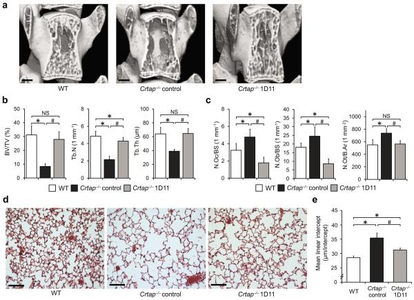
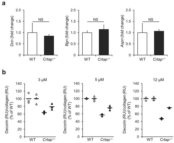
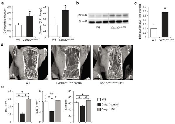
Osteogenesis imperfecta (OI) is a heritable disorder, in both a dominant and recessive manner, of connective tissue characterized by brittle bones, fractures and extraskeletal manifestations. How structural mutations of type I collagen (dominant OI) or of its post-translational modification machinery (recessive OI) can cause abnormal quality and quantity of bone is poorly understood. Notably, the clinical overlap between dominant and recessive forms of OI suggests common molecular pathomechanisms. Here, we show that excessive transforming growth factor-β (TGF-β) signaling is a mechanism of OI in both recessive (Crtap(-/-)) and dominant (Col1a2(tm1.1Mcbr)) OI mouse models. In the skeleton, we find higher expression of TGF-β target genes, higher ratio of phosphorylated Smad2 to total Smad2 protein and higher in vivo Smad2 reporter activity. Moreover, the type I collagen of Crtap(-/-) mice shows reduced binding to the small leucine-rich proteoglycan decorin, a known regulator of TGF-β activity. Anti-TGF-β treatment using the neutralizing antibody 1D11 corrects the bone phenotype in both forms of OI and improves the lung abnormalities in Crtap(-/-) mice. Hence, altered TGF-β matrix-cell signaling is a primary mechanism in the pathogenesis of OI and could be a promising target for the treatment of OI.


Figures

Figure 1
Figure 1
Excessive TGFβ signaling in Crtap−/− mice. (a) Quantitative RT-PCR of TGFβ target genes Cdkn1a and Serpine1 in calvarial bone of P3 WT and Crtap−/− mice. Results are shown as fold change of the mean of WT group±SD; n=5 per group. (b) Western blot analysis of P3 calvarial protein extracts showing amounts of activated Smad2 (pSmad2) relative to total Smad2 protein in Crtap−/− versus WT mice; n=3 per group. (c) Quantification of the Western blot shown in b. Results are shown as fold change of the mean of WT group±SD. (d) Bioluminescence in regions that overlap with skeletal structures in Crtap−/− compared with WT mice that were intercrossed to TGFβ-reporter mice (SBE-Luc mice). Representative image of 3 litters at P10 is shown (scale bar=1cm). (e) TGFβ activity in conditioned medium from WT and Crtap−/− bone marrow stromal cells cultured under osteogenic conditions. Results are shown as fold change of the mean of WT group±SD, n=6 per group. (f) Immunostaining of lungs (P10) for pSmad2 (red) in WT and Crtap−/− mice, DAPI (blue) staining of nuclei (40X magnification). Representative images of n=3 mice per group are shown (scale bar=20μm). *P<0.05 WT versus Crtap−/−.
Figure 2
Figure 2
Phenotypic correction of Crtap−/− mice after treatment with the TGFβ neutralizing antibody 1D11. (a) MicroCT images of L4 vertebral bodies of 16-week-old wildtype (WT), control antibody-treated Crtap−/−, and 1D11-treated Crtap−/− mice after treatment for 8 weeks (scale bar=500μm). (b) MicroCT analysis results of L4 vertebral bodies for bone volume/total volume (BV/TV), trabecular number (Tb.N) and trabecular thickness (Tb.Th) in WT, control Crtap−/− and 1D11 treated Crtap−/− mice. Results are shown as means±SDs, n=8 per group. (c) Histomorphometric analysis of L4 for osteoclast (N.Oc/BS) and osteoblast (N.Ob/BS) numbers per bone surface, and numbers of osteocytes per bone area (N.Ot/B.Ar) in WT, control Crtap−/− and 1D11 treated Crtap−/− mice. Results are shown as means±SDs, n=6 per group. (d) Hematoxylin/eosin staining of inflated lungs of 16-week-old wildtype (WT), control Crtap−/−, and 1D11-treated Crtap−/− mice after treatment for 8 weeks. Representative images of n=8 mice per group are shown (scale bar=100 μm). (e) Quantification of the distance between alveolar structures by the mean-linear-intercept (MLI) method in lungs of WT, control Crtap−/− and 1D11-treated Crtap−/− mice. Results are shown as means±SDs, n=8 mice per group, 10 images analyzed per mouse. *P<0.05 for Crtap−/− vs. WT, #P<0.05 for Crtap−/− 1D11 vs. Crtap−/− control. NS, not significant.
Figure 3
Figure 3
Reduced decorin binding to type I collagen of Crtap−/− mice. (a) Quantitative RTPCR of calvarial bone of P3 mice for the small leucine-rich proteoglycans decorin (Dcn), biglycan (Bgn), and asporin (Aspn) in WT and Crtap−/− mice. Results given as fold change of the mean of WT group±SD, n=5 per group. NS, not significant. (b) Surface plasmon resonance analysis measuring the binding of recombinant decorin core protein to type I collagen of WT and Crtap−/− mice. Three technical replicates at each of the indicated concentrations of decorin were performed in two independent biological replicates (◆ replicate 1, ▲ replicate 2). Results are shown as the percentage of the mean of WT (bars indicate mean per group).
Figure 4
Figure 4
Inhibition of upregulated TGFβ signaling improves the bone phenotype in a mouse model of dominant OI (Col1a2tm1.1Mcbr). (a) Quantitative RT-PCR of TGFβ target genes Cdkn1a and Serpine1 in calvarial bone of P3 WT and Col1a2tm1.1Mcbr mice. Results are shown as fold change of the mean of WT group±SD; n=3 per group. (b) Western blot analysis showing activated Smad2 (pSmad2) relative to total Smad2 protein in P3 calvaria of WT and Col1a2tm1.1Mcbr mice; n=3 per group. (c) Quantification of the Western blot seen in b. Results are shown as fold change of the mean of WT group±SD. (d) MicroCT images of L4 vertebral bodies of 16-week-old wildtype (WT), control antibody-treated Col1a2tm1.1Mcbr and 1D11-treated Col1a2tm1.1Mcbr mice after treatment for 8 weeks (scale bar=500 μm). (e) MicroCT analysis results of L4 vertebral bodies for bone volume/total volume (BV/TV), trabecular number (Tb.N) and thickness (Tb.Th) in WT, control Col1a2tm1.1Mcbr and 1D11 treated Col1a2tm1.1Mcbr mice. Results are shown as means±SDs, n=6 per group. *P<0.05 for Crtap−/− vs. WT, #P<0.05 for Crtap−/− 1D11 vs. Crtap−/− control. NS, not significant.

References

    1. Rauch F, Glorieux FH. Osteogenesis imperfecta. Lancet. 2004;363:1377–1385. - PubMed
    1. Baldridge D, et al. CRTAP and LEPRE1 mutations in recessive osteogenesis imperfecta. Hum Mutat. 2008;29:1435–1442. - PMC - PubMed
    1. Markmann A, Hausser H, Schonherr E, Kresse H. Influence of decorin expression on transforming growth factor-beta-mediated collagen gel retraction and biglycan induction. Matrix Biol. 2000;19:631–636. - PubMed
    1. Takeuchi Y, Kodama Y, Matsumoto T. Bone matrix decorin binds transforming growth factor-beta and enhances its bioactivity. J Biol Chem. 1994;269:32634–32638. - PubMed
    1. Morello R, et al. CRTAP is required for prolyl 3- hydroxylation and mutations cause recessive osteogenesis imperfecta. Cell. 2006;127:291–304. - PubMed

Publication types

MeSH terms