. 2014 Jun;35(6):716-26.
doi: 10.1038/aps.2013.203. Epub 2014 May 5.

Atorvastatin prevents amyloid-β peptide oligomer-induced synaptotoxicity and memory dysfunction in rats through a p38 MAPK-dependent pathway

Affiliations

Atorvastatin prevents amyloid-β peptide oligomer-induced synaptotoxicity and memory dysfunction in rats through a p38 MAPK-dependent pathway

Ling-Ling Zhang et al. Acta Pharmacol Sin. 2014 Jun.

Abstract

Aim: To investigate whether atorvastatin treatment could prevent Aβ1-42 oligomer (AβO)-induced synaptotoxicity and memory dysfunction in rats, and to elucidate the mechanisms involved in the neuroprotective actions of atorvastatin.

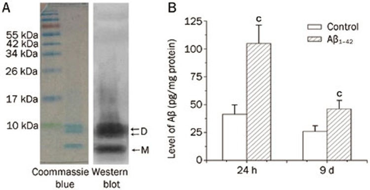
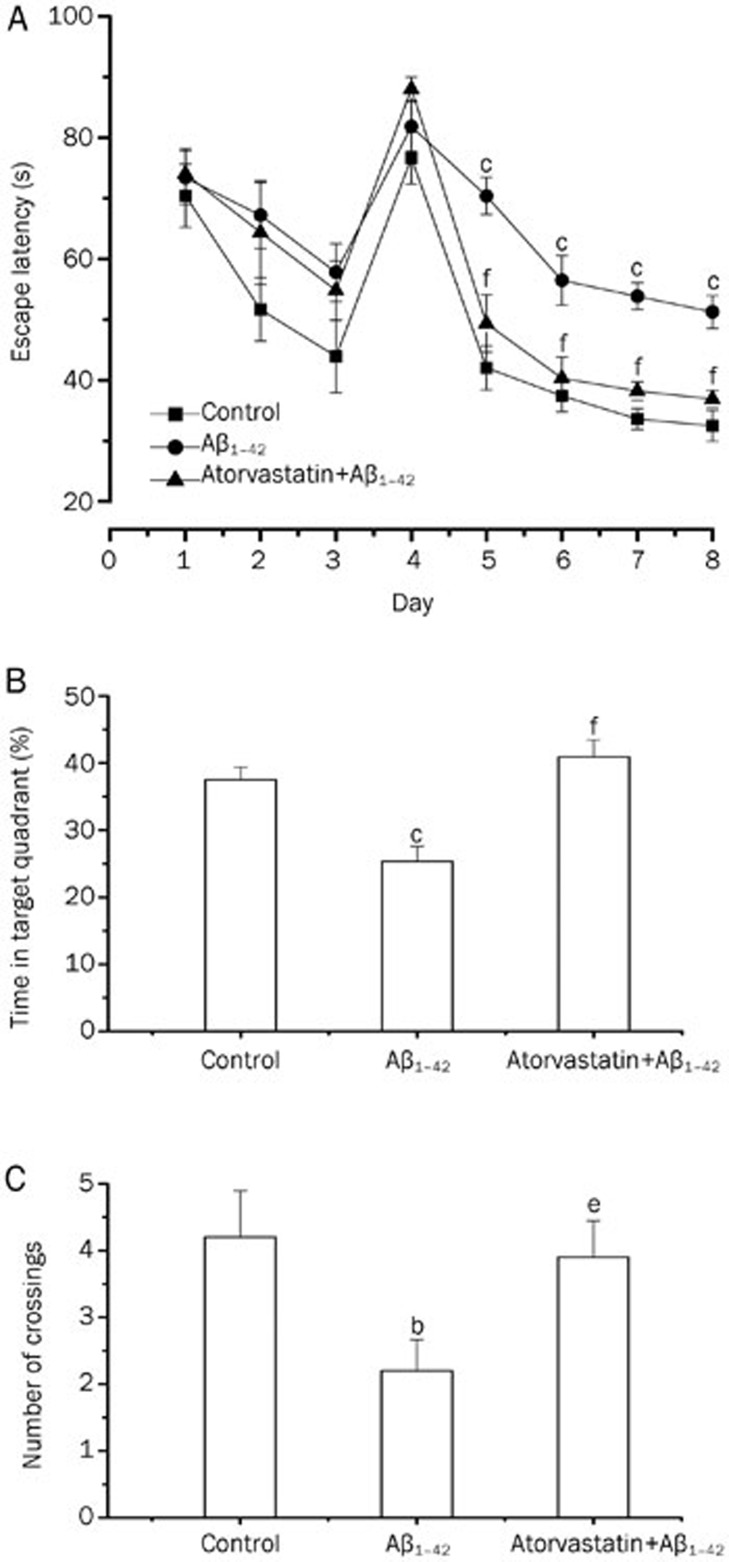
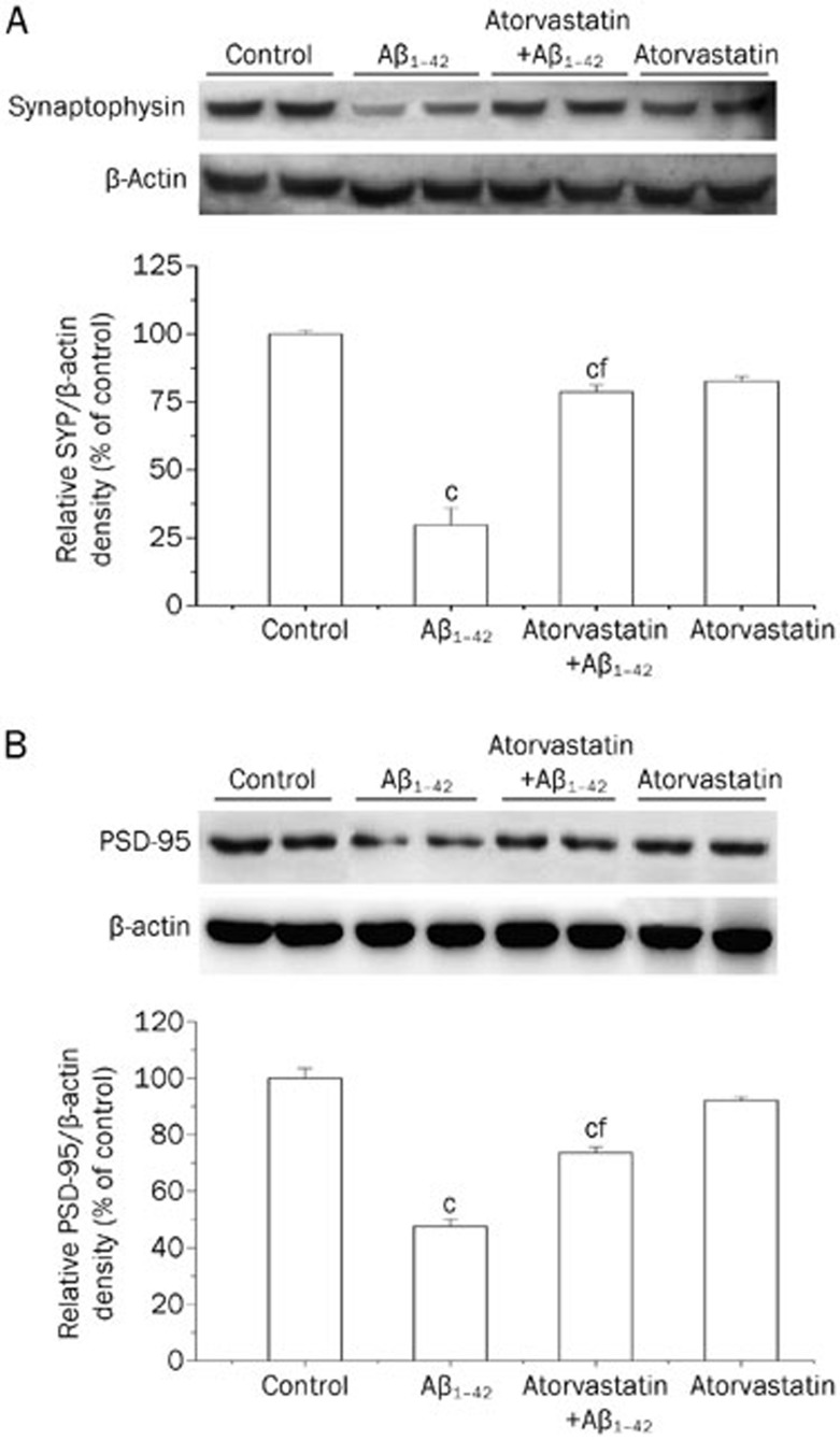
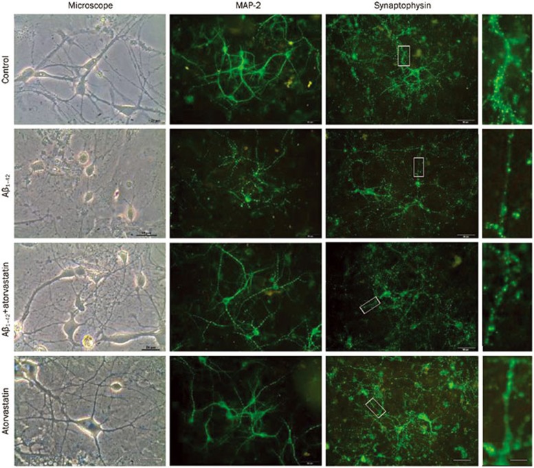
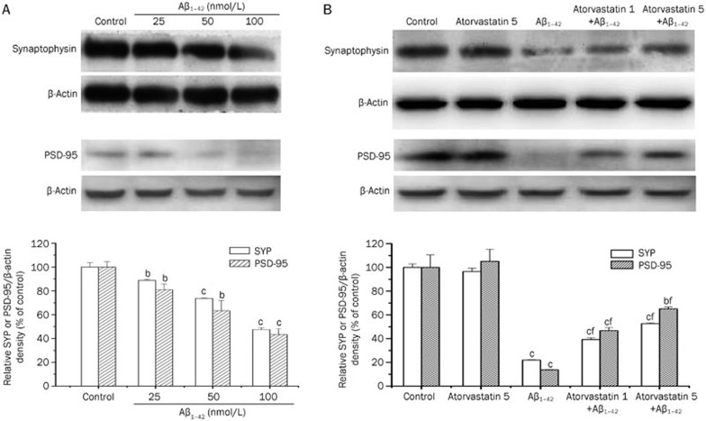
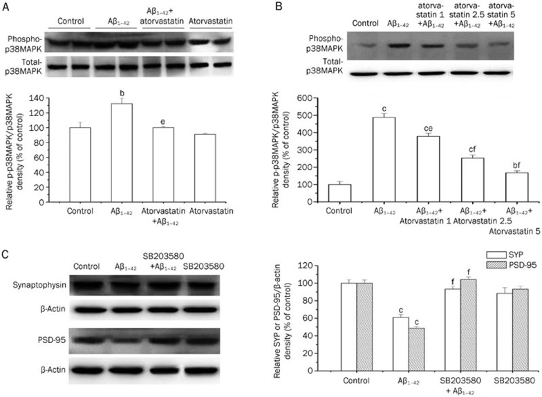
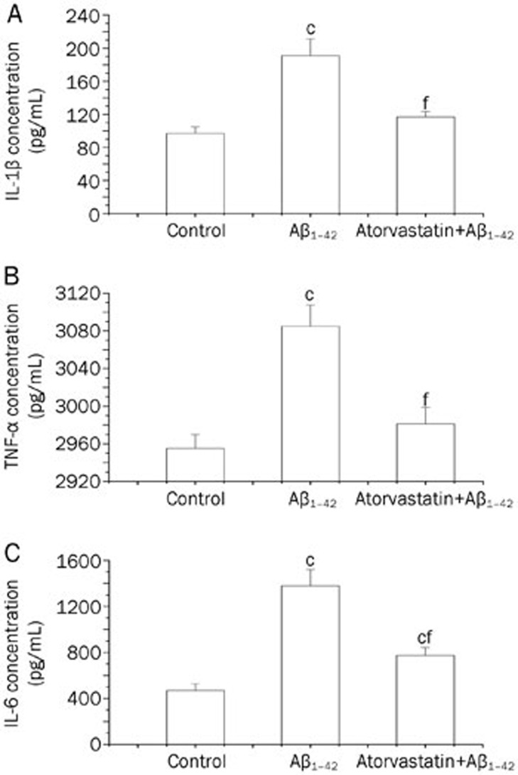
Methods: SD rats were injected with AβOs (5 nmol, icv). The rats were administrated with atorvastatin (10 mg·kg(-1)·d(-1), po) for 2 consecutive weeks (the first dose was given 5 d before AβOs injection). The memory impairments were evaluated with Morris water maze task. The expression of inflammatory cytokines in the hippocampus was determined using ELISA assays. The levels of PSD-95 and p38MAPK proteins in rat hippocampus were evaluated using Western blot analysis. For in vitro experiments, cultured rat hippocampal neurons were treated with AβOs (50 nmol/L) for 48 h. The expression of MAP-2 and synaptophysin in the neurons was detected with immunofluorescence.

Results: The AβO-treated rats displayed severe memory impairments in Morris water maze tests, and markedly reduced levels of synaptic proteins synaptophysin and PSD-95, increased levels of inflammatory cytokines (IL-1β, IL-6 and TNF-α) and p38MAPK activation in the hippocampus. All these effects were prevented or substantially attenuated by atorvastatin administration. Pretreatment of cultured hippocampal neurons with atorvastatin (1 and 5 μmol/L) concentration-dependently attenuated the AβO-induced synaptotoxicity, including the loss of dendritic marker MAP-2, and synaptic proteins synaptophysin and PSD-95. Pretreatment of the cultured hippocampal neurons with the p38MAPK inhibitor SB203580 (5 μmol/L) blocked the AβO-induced loss of synaptophysin and PSD-95.

Conclusion: Atorvastatin prevents AβO-induced synaptotoxicity and memory dysfunction through a p38MAPK-dependent pathway.


Figures

Figure 1
Figure 1
Identification of Aβ peptide solutions and accumulation of Aβ in the hippocampus after icv administration. (A) Coomassie brilliant blue-stained SDS-PAGE gel image and anti-antibody-based Western blot analysis of Aβ1–42 showed that the solutions primarily consisted of dimers and monomers. The positions of Aβ dimmers (D) and monomers (M) are indicated by arrows on the right side of the blot. (B) Aβ1–42 accumulated in the hippocampus 24 h and 9 d after icv administration, as measured by ELISA. Data are expressed as the mean±SEM. n=5. cP<0.01 compared with the control group.
Figure 2
Figure 2
Atorvastatin prevented AβO-induced learning and memory deficits in rats. AβO-treated rats displayed longer latencies to reach the hidden platform (d 5–8) (A), as well as decreased time (B) and number of platform crossings (C) in the target quadrant during the probe trial compared with the control rats. Data are expressed as the mean±SEM of 12 rats. bP<0.05, cP<0.01 compared with the control group. eP<0.05, fP<0.01 compared with the AβO-treated group.
Figure 3
Figure 3
Western blot analysis showing the effects of atorvastatin on the AβO-induced decrease in synaptophysin and PSD-95 protein expression in rat hippocampus. Rats were treated with AβOs (5 nmol, icv) or sterile water (control). Atorvastatin (10 mg/kg) was administered daily (starting from 5 d before AβO treatment). (A) Synaptophysin protein levels. (B) PSD-95 protein levels. The bar chart shows the semiquantitative analysis of synaptophysin and PSD-95. Data are expressed as the mean±SEM of 4 independent preparations. cP<0.01 compared with the control group. fP<0.01 compared with the AβO-treated group.
Figure 4
Figure 4
Representative images of cultured hippocampal neurons obtained via a phase-contrast microscope (scale bar, 25 μm) and immunolabeled against MAP-2 or synaptophysin (scale bar, 50 μm). Insets on the right side represent digital enlargements (scale bar, 200 μm) of the dendrite segments indicated by white boxes. Cultured hippocampal neurons at d 7 were pretreated with vehicle solution (control, 0.1% DMSO) or atorvastatin (5 μmol/L) for 1 h and then exposed to AβOs (50 nmol/L) for 48 h in the presence of vehicle or atorvastatin. Following the treatment period, phase-contrast digital images of the neurons were taken using a phase-contrast microscope. Hippocampal neurons were immunostained for MAP-2 or synaptophysin after 48 h incubation with AβOs (50 nmol/L) and analyzed by confocal microscopy.
Figure 5
Figure 5
Atorvastatin prevented the decrease in AβO-induced synaptophysin and PSD-95 in cultured hippocampul neurons. (A) Representative Western blot of synaptophysin and PSD-95. Group data showing the normalization of synaptophysin and PSD-95 proteins to β-actin protein was determined in each group from 4 experiments. (B) Representative Western blot showing that atorvastatin (1 and 5 μmol/L) prevented AβO-induced decreases in synaptophysin and PSD-95 in a concentration-dependent manner. Group data showing the normalization of synaptophysin and PSD-95 proteins to β-actin protein was determined in each group from 4 experiments. Data are expressed as the mean±SEM. bP<0.05, cP<0.01 compared with the control group. fP<0.01 compared with the AβO-treated group.
Figure 6
Figure 6
The neuroprotection afforded by atorvastatin against AβO-induced synaptotoxicity involved the p38MAPK signaling pathway. (A) Representative Western blot comparing phospho-p38MAPK in rat hippocampus. Group data showing the normalization of phospho-p38MAPK to total p38MAPK was determined in each group from 4 experiments. (B) Representative Western blot comparing phospho-p38MAPK in cultured hippocampal neurons. Cultured hippocampal neurons at d 7 were pre-incubated with vehicle solution (control, 0.1% DMSO) or atorvastatin (1, 2.5, or 5 μmol/L) for 1 h and then exposed to AβOs (50 nmol/L) for 12 h in the presence of vehicle or atorvastatin. Group data showing the normalization of phospho-p38MAPK to total p38MAPK were determined in each group from 4 experiments. (C) Representative Western blot showing that the p38MAPK inhibitor SB203580 prevented the decrease of synaptophysin and PSD-95 proteins induced by AβOs. Hippocampal neurons were pre-incubated with vehicle solution (control, 0.1% DMSO) or SB203580 (5 μmol/L) for 30 min and then exposed to AβOs (50 nmol/L) for 48 h in the presence of vehicle or SB203580. Group data showing the normalization of synaptophysin and PSD-95 proteins to β-actin protein were determined in each group from 4 experiments. Data are expressed as the mean±SEM. bP<0.05, cP<0.01 compared with the control group. eP<0.05, fP<0.01 compared with the AβO-treated group.
Figure 7
Figure 7
Atorvastatin suppressed the proinflammatory cytokine upregulation induced by AβOs. Treatment with atorvastatin resulted in a significant suppression of the AβO-induced increase in the hippocampal levels of the proinflammatory cytokines IL-1β (A), TNF-α (B), and IL-6 (C). Rats were treated with AβOs (5 nmol, icv) or sterile water (control). Atorvastatin (10 mg/kg) was administered daily starting 5 d before AβO treatment. Rats were euthanized on d 9, and hippocampal extracts were analyzed by ELISA. Data are expressed as the mean±SEM of 5 rats per group. cP<0.01 compared with the control group. fP<0.01 compared with the AβO-treated group.

Similar articles

Cited by

References

    1. Haass C, Selkoe DJ. Soluble protein oligomers in neurodegeneration: lessons from the Alzheimer's amyloid beta-peptide. Nat Rev Mol Cell Biol. 2007;8:101–12. - PubMed
    1. Cerpa W, Dinamarca MC, Inestrosa NC. Structure–function implications in Alzheimer's disease: effect of Abeta oligomers at central synapses. Curr Alzheimer Res. 2008;5:233–43. - PubMed
    1. Gong Y, Chang L, Viola KL, Lacor PN, Lambert MP, Finch CE, et al. Alzheimer's disease-affected brain: presence of oligomeric A beta ligands (ADDLs) suggests a molecular basis for reversible memory loss. Proc Natl Acad Sci U S A. 2003;100:10417–22. - PMC - PubMed
    1. Chang L, Bakhos L, Wang Z, Venton DL, Klein WL. Femtomole immunodetection of synthetic and endogenous amyloid-beta oligomers and its application to Alzheimer's disease drug candidate screening. J Mol Neurosci. 2003;20:305–13. - PubMed
    1. Cleary JP, Walsh DM, Hofmeister JJ, Shankar GM, Kuskowski MA, Selkoe DJ, et al. Natural oligomers of the amyloid-beta protein specifically disrupt cognitive function. Nat Neurosci. 2005;8:79–84. - PubMed

Publication types

MeSH terms