. 2014 Jun;13(6):631-7.
doi: 10.1038/nmat3960. Epub 2014 May 4.

Rigidity sensing and adaptation through regulation of integrin types

Affiliations

Rigidity sensing and adaptation through regulation of integrin types

Alberto Elosegui-Artola et al. Nat Mater. 2014 Jun.

Abstract

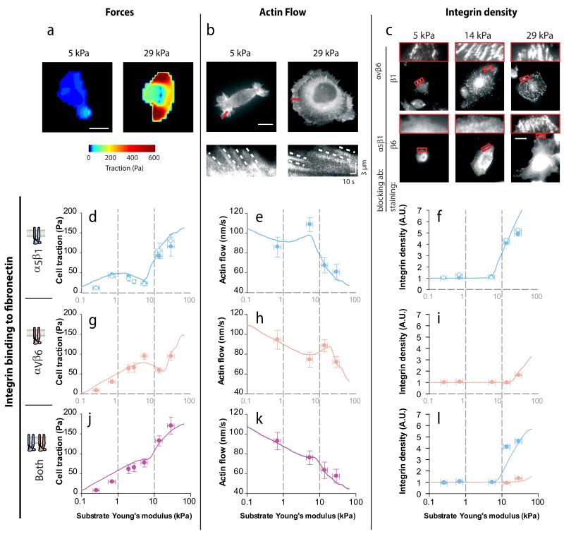
Tissue rigidity regulates processes in development, cancer and wound healing. However, how cells detect rigidity, and thereby modulate their behaviour, remains unknown. Here, we show that sensing and adaptation to matrix rigidity in breast myoepithelial cells is determined by the bond dynamics of different integrin types. Cell binding to fibronectin through either α5β1 integrins (constitutively expressed) or αvβ6 integrins (selectively expressed in cancer and development) adapts force generation, actin flow and integrin recruitment to rigidities associated with healthy or malignant tissue, respectively. In vitro experiments and theoretical modelling further demonstrate that this behaviour is explained by the different binding and unbinding rates of both integrin types to fibronectin. Moreover, rigidity sensing through differences in integrin bond dynamics applies both when integrins bind separately and when they compete for binding to fibronectin.


Figures

Figure 1
Figure 1. Expression of αvβ6 integrins alters response to substrate stiffness
(a) Colour maps showing the traction forces applied by individual myo ctrl or myo β6 cells on FN-coated polyacrylamide gels of increasing rigidity. (b) Average forces as a function of rigidity for myo ctrl cells (red) and myo β6 cells (blue). (c) Average forces for both cell types after blocking αvβ6 with an inhibitory antibody. Differences between cell types were significant without antibodies (p<0.05) but not with antibodies, and the effect of stiffness was significant in all cases (p<0.05). n≥11 cells per condition. Scale bar is 20 μm.
Figure 2
Figure 2. Both binding and unbinding rates to FN are higher for αvβ6 than for α5β1
(a) Surface Plasmon Resonance curves showing attachment and subsequent detachment of solutions of purified α5β1 integrins and αvβ6 integrins to a FN-coated substrate. Lines of increasing height represent increasing integrin concentrations (9-264 nM for α5β1 and 67-527 nM for αvβ6). Data for 264 nM is marked in bold in both cases. Fitted kont and koff values are shown below graph (n ≥3 experiments). (b) Integrin densities on the membrane for both cell types (n≥46 cells per condition, see also Supplementary Figure 5). (c) Top: The tip of a magnetic tweezers device was approached to cells with attached FN-coated magnetic beads, and used to apply a force of 0.5 nN to beads for 2 minutes. Scale bar is 20 μm. Bottom: time required to detach beads from myo ctrl or myo β6 cells (with or without blocking antibodies) after force application (n≥64 beads from ≥47 cells per condition). (d) DIC images and β1 integrin staining images of myo ctrl or myo β6 cells (with or without blocking αvβ6 integrins) showing β1 integrin recruitment to FN-coated beads. Insets show beads marked with a red square. (e) Corresponding quantification of integrin recruitment to beads (n≥20 beads from ≥10 cells per condition). (f-g) Same as d-e, but staining for β6 integrins and blocking α5β1 integrins instead of the reverse (n≥ 23 beads from ≥ 15 cells per condition). Scale bar is 20 μm. *: p<0.05.
Figure 3
Figure 3. Integrin-FN clutch model of force transmission
(a) Myosin motors pull on actin filaments, which move with rearward speed va. Integrins of two different types connect to the actin flow through adaptor proteins, and compete for binding to FN with effective binding rates kon1 and kon2 given by true binding rates (kont1, kont2) multiplied by integrin densities on the membrane (dint1, dint2). FN molecules are in turn connected to a compliant substrate, represented as a linear elastic spring of varying rigidity. (b) Flow of events (from top to bottom). (i) The model considers a given number of FN molecules (triangles) attached to the substrate, to which integrins can bind. (ii) Orange integrins, with higher kon, will bind faster. (iii) Once they bind, actin rearward movement applies a force on all integrins and the substrate. Because orange integrins also have a higher koff, they will detach sooner. (iv) Eventually, blue integrins will detach as well, and if before detaching the force transmitted through them reaches a threshold value, a reinforcement mechanosensing event will result in increased integrin density. (v) Once all integrins detach, force on the substrate is released and the cycle begins again. (c-e) Cell traction forces (c), integrin densities (d), and actin speeds (e) predicted by the model, the three regimes (1-3) are discussed in the main text and in Supplementary Note 4.
Figure 4
Figure 4. Integrin-fibronectin binding dynamics predict force generation, actin flow, and integrin recruitment in response to substrate stiffness
(a) Examples of traction maps exerted by myo ctrl cells plated on substrates of 5 or 29 kPa. (b) Top: examples of myo ctrl cells transfected with lifeact-GFP plated on substrates of 5 or 29 kPa. Bottom: kymographs showing the movement of actin features along the lines marked in red in the top image. The slope of the traces created by the features (marked with dotted lines) was used to calculate actin speed. (c) Staining of β1 integrins in myo β6 cells where αvβ6 was blocked, and of β6 integrins in cells where α5β1 was blocked. Zoomed regions on top of images correspond to rectangles marked in red in the main image. Scale bars are 20 μm. (d-l) For cells on FN-coated gels of varying stiffness, quantifications of average cell traction forces (d,g,j, n≥14 cells per condition), actin rearward flows (e,h,k, n≥19 traces from ≥7 cells per condition), and integrin densities in adhesions (f,I,l, n≥52 adhesions from ≥10 cells per condition). Results are shown for cells with adhesion mediated by α5β1 integrins (d-f; ●: myo ctrl, ○: myo β6 + αvβ6 ab), αvβ6 integrins (g-i, myo β6+ α5β1 ab), and both integrins (j-l, myo β6 cells without any ab). Lines indicate model predictions. In panel l, blue and orange symbols correspond respectively to β1 and β6 integrin densities. Statistical analyses are detailed in methods.

Comment in

References

    1. Wang HB, Dembo M, Wang YL. Substrate flexibility regulates growth and apoptosis of normal but not transformed cells. Am J Physiol Cell Physiol. 2000;279(5):C1345–1350. - PubMed
    1. Engler AJ, Sen S, Sweeney HL, Discher DE. Matrix elasticity directs stem cell lineage specification. Cell. 2006;126(4):677–689. - PubMed
    1. Paszek MJ, Zahir N, Johnson KR, Lakins JN, Rozenberg GI, Gefen A, et al. Tensional homeostasis and the malignant phenotype. Cancer Cell. 2005;8(3):241–254. - PubMed
    1. Ghassemi S, Meacci G, Liu S, Gondarenko AA, Mathur A, Roca-Cusachs P, et al. Cells test substrate rigidity by local contractions on sub-micrometer pillars. PNAS. 2012;109(14):5328–5333. - PMC - PubMed
    1. Trichet L, Le Digabel J, Hawkins RJ, Vedula SR, Gupta M, Ribrault C, et al. Evidence of a large-scale mechanosensing mechanism for cellular adaptation to substrate stiffness. PNAS. 2012;109(18):6933–6938. - PMC - PubMed

Publication types