Review
. 2014 Jul;34 Suppl 1(0 1):S35-45.
doi: 10.1007/s10875-014-0022-7. Epub 2014 May 3.

The long elusive IgM Fc receptor, FcμR

Affiliations
Review

The long elusive IgM Fc receptor, FcμR

Hiromi Kubagawa et al. J Clin Immunol. 2014 Jul.

Abstract

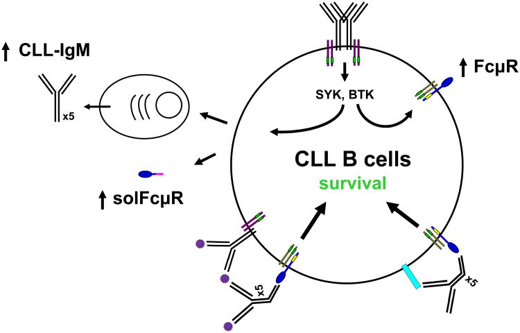
IgM exists as both a monomer on the surface of B cells and a pentamer secreted by plasma cells. Both pre-immune "natural" and antigen-induced "immune" IgM antibodies are important for protective immunity and for immune regulation of autoimmune processes by recognizing pathogens and self-antigens. Effector proteins interacting with the Fc portion of IgM, such as complement and complement receptors, have thus far been proposed but fail to fully account for the IgM-mediated protection and regulation. A major reason for this deficit in our understanding of IgM function seems to be lack of data on a long elusive Fc receptor for IgM (FcμR). We have recently identified a bona fide FcμR in both humans and mice. In this article we briefly review what we have learned so far about FcμR.


Conflict of interest statement

Conflict of interest disclosure: The authors declare no conflicting financial interests

Figures

Figure 1
Figure 1
Predicted protein structure of human FcμR. The human FcμR cDNA encodes a type I transmembrane protein of 390 aa and with a peptide core of ∼41 kDa. Numbers indicate aa residues in each region: signal peptide (SP), a single Ig-like domain (V-set), remaining extracellular (stalk), transmembrane (TM; between the two lines) and cytoplasmic region. Hatch marks indicate exon boundaries in the FCMR gene. A small closed circle within the transmembrane segment indicates a charged histidine residue (His).
Figure 2
Figure 2
Schematic chromosomal localization of FCMR. Partial chromosome 1 linkage map showing a cluster of three IgM-binding receptors (FCMR, PIGR, FCAMR) in 1q32 in relation to other FcR genes in 1q21 – 1q23.
Figure 3
Figure 3
Amino acid sequence alignment of IgM-binding receptors. The Ig-binding domains of pIgR, Fcα/μR and FcμR from several species are aligned with each other. Amino acid identity is indicated by dots (·) and a deletion by slashes (/). Residues conserved in all three receptors and in pIgR and Fcα/μR are highlighted in yellow and red, respectively. Accession codes for these sequences are: pIgR of human (P01833), rabbit (P01832), mouse (070570), rat (P15083), bovine (P81265), and chicken (AAP69798); Fcα/μR of human (AAL51154) and mouse (NP_659209); and FcμR of human (NP_005440), mouse (NP_081252), and rat (Q5M871). Crystallographically determined secondary structure elements and the topology diagram of human pIgR, which are determined by Hamburger et al., are shown above the sequences and in a right lower corner, respectively. The β strands A, B, E, and D are shown in blue, β strands C″, C′, C, F, G, and A′ are in green, and three CDR loops (including the α helix within CDR1) are red. The permission to incorporate the structural data has been obtained from Dr. Pamela Bjorkman.
Figure 4
Figure 4
Effect of IgM exposure and PMA treatment on the expression of IgM receptor by the 697 pre-B cell line. Cells were incubated for 16 h at 37°C with mouse IgM (0.3 mg/ml; top panel) or PMA (10 nM; bottom panel), washed, then assessed for IgM binding by flow cytometric analysis using biotin-labeled, rat anti-mouse μ mAb (left column), human IgM (middle column), mouse anti-human FcμR or isotype-matched control mAb (right column). The bound biotin-labeled reagents were detected by addition of phycoerythrin-labeled streptavidin (PE-SA). In the left two columns, the red lines are the reactivity of the indicated biotin reagents to cells preincubated with mouse IgM or PMA and the shaded histograms are that to cells preincubated with medium only as controls. In the right column, the red lines and shaded histograms are the reactivity of cells with anti-FcμR or isotype-matched control mAb, respectively. Note that mouse IgM-exposed 697 pre-B cells display already-bound mouse IgM and minimal binding of human IgM, but are negative for FcμR. By contrast, PMA-treated 697 pre-B cells clearly exhibit IgM binding and are positive for FcμR.
Figure 5
Figure 5
Amino acid sequence alignment of the transmembrane and cytoplasmic regions of FcμRs. The transmembrane and cytoplasmic regions of FcμR from six species are aligned with each other. Amino acid identity is indicated by dots (·) and a deletion by dashes (-). The predicted transmembrane region is colored in pink. Conserved Tyr, Ser and Cys residues are also highlighted in yellow, dark or light blue, and purple, respectively. Light blue indicate conservation of Ser residues in five species. The numbers indicate the aa position from the first Met residue of human FcμR.
Figure 6
Figure 6
FcμR expression by B cell subsets in blood and tonsils. MNCs from adult blood (A) and tonsils (B) were first incubated with FcγR-blocking reagents and then with biotin-labeled, anti-FcμR (HM14; γ1κ) or isotype-matched control mAb, before developing with PE-SA. PE-stained cells were counterstained with fluorochrome-labeled mixture of four or three mAbs with specificity for: CD19, CD20, CD21 or CD27 (A and B lower panel) or CD19, IgD or CD38 (B upper panel), including fluorochrome-labeled, corresponding isotype-matched control mAbs for background setting. Stained cells were analyzed by BD LSR II (A and B lower) and Accuri C6 (B upper) flow cytometries. Cells in boxes with numbers or different color frames were examined for the reactivity with FcμR-specific (solid lines) or control (shaded histograms) mAb. Numbers indicate the mean fluorescence intensity (MFI) ratios defined as (MFI of anti-FcμR mAb ÷ MFI of control mAb). AM, activated memory; TLM, tissue-like memory; RM, resting memory; FO, follicular; preGC, pregerminal center; Mem, memory; GC, germinal center; PC, plasma cell.
Figure 6
Figure 6
FcμR expression by B cell subsets in blood and tonsils. MNCs from adult blood (A) and tonsils (B) were first incubated with FcγR-blocking reagents and then with biotin-labeled, anti-FcμR (HM14; γ1κ) or isotype-matched control mAb, before developing with PE-SA. PE-stained cells were counterstained with fluorochrome-labeled mixture of four or three mAbs with specificity for: CD19, CD20, CD21 or CD27 (A and B lower panel) or CD19, IgD or CD38 (B upper panel), including fluorochrome-labeled, corresponding isotype-matched control mAbs for background setting. Stained cells were analyzed by BD LSR II (A and B lower) and Accuri C6 (B upper) flow cytometries. Cells in boxes with numbers or different color frames were examined for the reactivity with FcμR-specific (solid lines) or control (shaded histograms) mAb. Numbers indicate the mean fluorescence intensity (MFI) ratios defined as (MFI of anti-FcμR mAb ÷ MFI of control mAb). AM, activated memory; TLM, tissue-like memory; RM, resting memory; FO, follicular; preGC, pregerminal center; Mem, memory; GC, germinal center; PC, plasma cell.
Figure 7
Figure 7
FcμR expression by T cell subsets in blood and tonsils. MNCs from blood (A) and tonsils (B) were similarly stained as in Fig. 6 for FcμR and counterstained with fluorochrome-labeled anti-CD4 (upper) or anti-CD8 (lower) mAb, along with other fluorochrome-labeled mAbs specific for CD45RA, CD27 or CCR7. Stained cells were similarly analyzed by BD LSR II flow cytometry as described in the Fig. 6 legend. EM, effector memory; CM, central memory; EF, effector; EMRA, effector memory CD45RA+.
Figure 7
Figure 7
FcμR expression by T cell subsets in blood and tonsils. MNCs from blood (A) and tonsils (B) were similarly stained as in Fig. 6 for FcμR and counterstained with fluorochrome-labeled anti-CD4 (upper) or anti-CD8 (lower) mAb, along with other fluorochrome-labeled mAbs specific for CD45RA, CD27 or CCR7. Stained cells were similarly analyzed by BD LSR II flow cytometry as described in the Fig. 6 legend. EM, effector memory; CM, central memory; EF, effector; EMRA, effector memory CD45RA+.
Figure 8
Figure 8
Hypothetical role of Fc μR in CLL. Our current working hypothesis of the role of FcμR in CLL is as follows. Antigen-independent self-ligation of BCR on CLL cells activates SYK and BTK tyrosine kinases and induces up-regulation of the cell surface expression of FcμR/adaptor protein complex. Subpopulations of CLL cells differentiate into plasma cells that secrete pentameric IgM antibodies. Secreted IgM antibodies recognize soluble (purple spiky small circles) or lymphocyte membrane (light blue rectangle) self-antigens. The colligation of FcμR and BCR by soluble IgM/self-antigen immune complexes or the cis interaction of FcμR and lymphocyte membrane self-antigen by secreted IgM provides a survival signal to CLL cells through FcμR. BCR is depicted as black Y shape heavy and light chain lines with Igα/β adaptor proteins (purple lines) carrying ITAM (small green rectangles). FcμR ligand-binding chain is depicted as a blue tennis racket shape with a small yellow rectangle indicating three conserved intracytoplasmic Tyr residues and associates with an unknown adaptor protein (gray lines) possibly carrying ITAM (small green rectangles). Soluble FcμR, an alternative splice variant, is also markedly elevated in CLL patients, but its biological function remains unknown.

Similar articles

Cited by

References

    1. Ravetch JV, Kinet JP. Fc receptors. Annu Rev Immunol. 1991;9:457–92. - PubMed
    1. Daëron M. Fc receptor biology. Annu Rev Immunol. 1997;15:203–34. - PubMed
    1. Monteiro RC, Van De Winkel JG. IgA Fc receptors. Annu Rev Immunol. 2003;21:177–204. - PubMed
    1. Nimmerjahn F, Bruhns P, Horiuchi K, Ravetch JV. FcγRIV: a novel FcR with distinct IgG subclass specificity. Immunity. 2005 Jul;23(1):41–51. - PubMed
    1. Kubagawa H, Oka S, Kubagawa Y, Torii I, Takayama E, Kang DW, et al. Identity of the elusive IgM Fc receptor (FcμR) in humans. J Exp Med. 2009 Nov 23;206(12):2779–93. - PMC - PubMed

Publication types