

Expression of T-cell KV1.3 potassium channel correlates with pro-inflammatory cytokines and disease activity in ulcerative colitis
- PMID: 24793818
- PMCID: PMC4216648
- DOI: 10.1016/j.crohns.2014.04.003
Expression of T-cell KV1.3 potassium channel correlates with pro-inflammatory cytokines and disease activity in ulcerative colitis
Abstract
Background and aims: Potassium channels, KV1.3 and KCa3.1, have been suggested to control T-cell activation, proliferation, and cytokine production and may thus constitute targets for anti-inflammatory therapy. Ulcerative colitis (UC) is a chronic inflammatory bowel disease characterized by excessive T-cell infiltration and cytokine production. It is unknown if KV1.3 and KCa3.1 in the inflamed mucosa are markers of active UC. We hypothesized that KV1.3 and KCa3.1 correlate with disease activity and cytokine production in patients with UC.
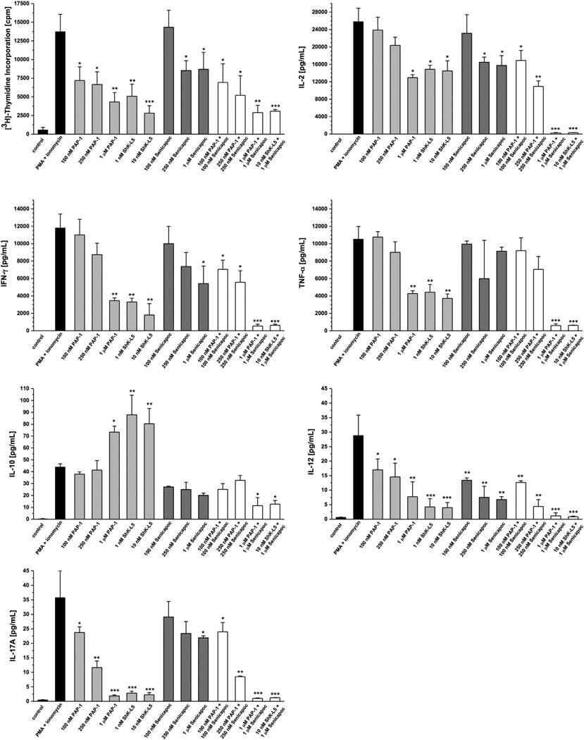
Methods: Mucosal biopsies were collected from patients with active UC (n=33) and controls (n=15). Protein and mRNA expression of KV1.3 and KCa3.1, immune cell markers, and pro-inflammatory cytokines were determined by quantitative-real-time-polymerase-chain-reaction (qPCR) and immunofluorescence, and correlated with clinical parameters of inflammation. In-vitro cytokine production was measured in human CD3(+) T-cells after pharmacological blockade of KV1.3 and KCa3.1.
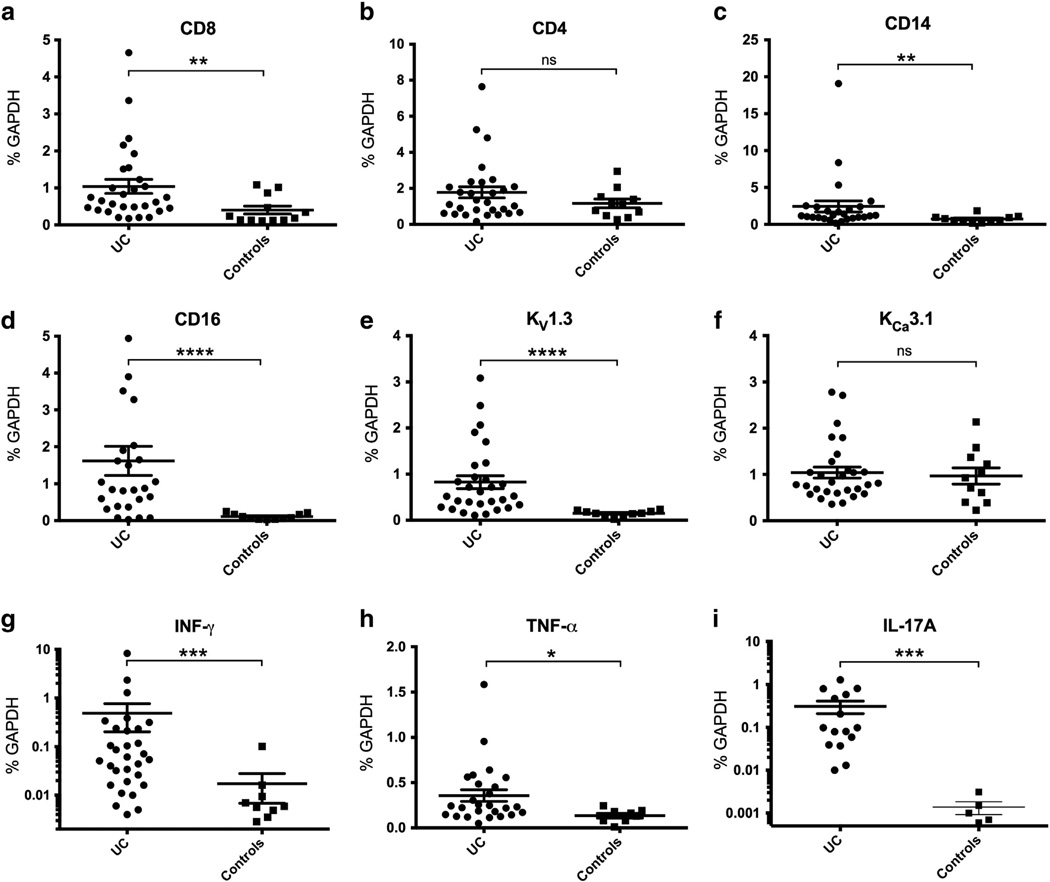
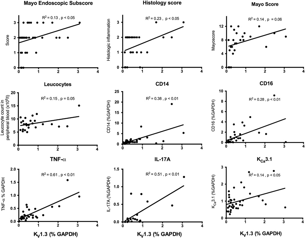
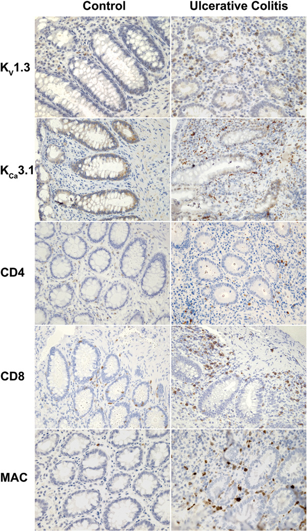
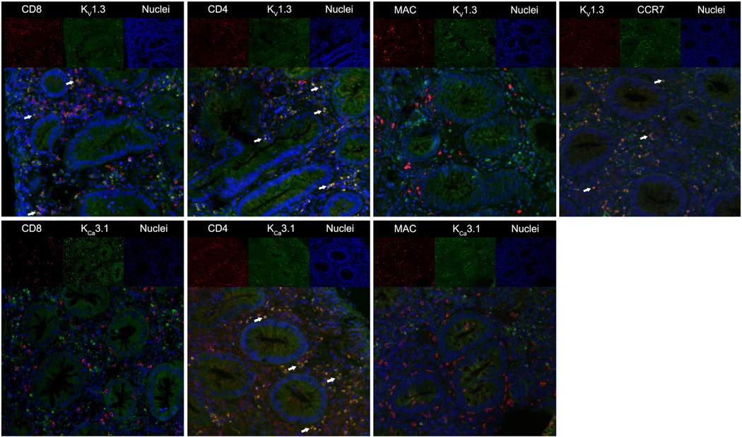
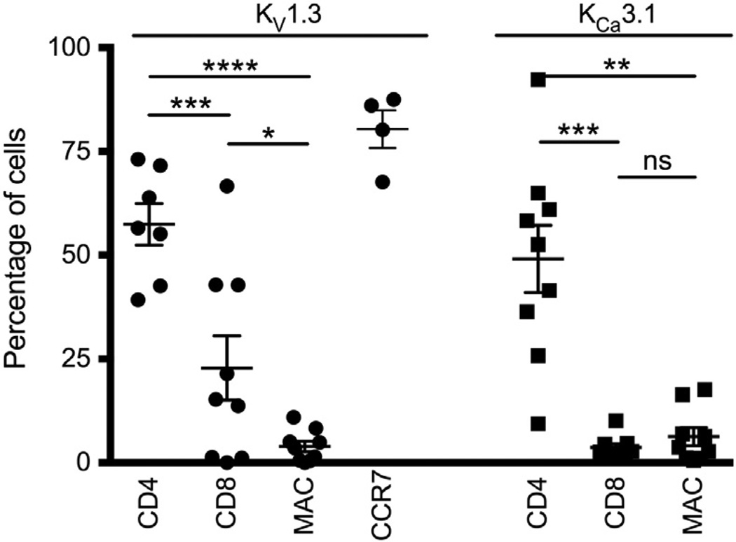
Results: Active UC KV1.3 mRNA expression was increased 5-fold compared to controls. Immunofluorescence analyses revealed that KV1.3 protein was present in inflamed mucosa in 57% of CD4(+) and 23% of CD8(+) T-cells. KV1.3 was virtually absent on infiltrating macrophages. KV1.3 mRNA expression correlated significantly with mRNA expression of pro-inflammatory cytokines TNF-α (R(2)=0.61) and IL-17A (R(2)=0.51), the mayo endoscopic subscore (R(2)=0.13), and histological inflammation (R(2)=0.23). In-vitro blockade of T-cell KV1.3 and KCa3.1 decreased production of IFN-γ, TNF-α, and IL-17A.
Conclusions: High levels of KV1.3 in CD4 and CD8 positive T-cells infiltrates are associated with production of pro-inflammatory IL-17A and TNF-α in active UC. KV1.3 may serve as a marker of disease activity and pharmacological blockade might constitute a novel immunosuppressive strategy.
Keywords: Colitis ulcerosa;; Interleukins;; K(Ca)3.1; KCNA3;; KCNN4;; Novel treatment strategy;.
Copyright © 2014 European Crohn's and Colitis Organisation. All rights reserved.
Conflict of interest statement
None.
Figures
Similar articles
-
The role of T cell potassium channels, KV1.3 and KCa3.1, in the inflammatory cascade in ulcerative colitis.Dan Med J. 2014 Nov;61(11):B4946. Dan Med J. 2014. PMID: 25370966 Review.
-
Inhibitory effects of candesartan on KCa3.1 potassium channel expression and cell culture and proliferation in peripheral blood CD4+T lymphocytes in Kazakh patients with hypertension from the Xinjiang region.Clin Exp Hypertens. 2018;40(4):303-311. doi: 10.1080/10641963.2017.1377212. Epub 2018 Feb 1. Clin Exp Hypertens. 2018. PMID: 29388859
-
Over-expression of interleukin 10 in mucosal T cells of patients with active ulcerative colitis.Clin Exp Immunol. 2003 Oct;134(1):127-37. doi: 10.1046/j.1365-2249.2003.02268.x. Clin Exp Immunol. 2003. PMID: 12974765 Free PMC article.
-
Quantitative gene expression of cytokines in peripheral blood leukocytes stimulated in vitro: modulation by the anti-tumor nerosis factor-alpha antibody infliximab and comparison with the mucosal cytokine expression in patients with ulcerative colitis.Transl Res. 2007 Oct;150(4):223-32. doi: 10.1016/j.trsl.2007.04.004. Epub 2007 May 11. Transl Res. 2007. PMID: 17900510
-
Development, validation and implementation of an in vitro model for the study of metabolic and immune function in normal and inflamed human colonic epithelium.Dan Med J. 2015 Jan;62(1):B4973. Dan Med J. 2015. PMID: 25557335 Review.
Cited by
-
Midazolam's Effects on Delayed-Rectifier K+ Current and Intermediate-Conductance Ca2+-Activated K+ Channel in Jurkat T-lymphocytes.Int J Mol Sci. 2021 Jul 4;22(13):7198. doi: 10.3390/ijms22137198. Int J Mol Sci. 2021. PMID: 34281255 Free PMC article.
-
Conditional KCa3.1-transgene induction in murine skin produces pruritic eczematous dermatitis with severe epidermal hyperplasia and hyperkeratosis.PLoS One. 2020 Mar 9;15(3):e0222619. doi: 10.1371/journal.pone.0222619. eCollection 2020. PLoS One. 2020. PMID: 32150577 Free PMC article.
-
N-Terminally extended analogues of the K⁺ channel toxin from Stichodactyla helianthus as potent and selective blockers of the voltage-gated potassium channel Kv1.3.FEBS J. 2015 Jun;282(12):2247-59. doi: 10.1111/febs.13294. Epub 2015 Apr 23. FEBS J. 2015. PMID: 25864722 Free PMC article.
-
Qingchi San treats ulcerative colitis in mice by inhibiting the nuclear factor-kappa B signaling pathway and Nucleotide-binding oligomerization domain, leucine-rich repeat and pyrin domain-containing 3 inflammasome formation.J Tradit Chin Med. 2023 Feb;43(1):68-77. doi: 10.19852/j.cnki.jtcm.20220928.001. J Tradit Chin Med. 2023. PMID: 36639997 Free PMC article.
-
Usefulness of targeting lymphocyte Kv1.3-channels in the treatment of respiratory diseases.Inflamm Res. 2015 Oct;64(10):753-65. doi: 10.1007/s00011-015-0855-4. Epub 2015 Jul 24. Inflamm Res. 2015. PMID: 26206235
References
-
- Sheth SG, LaMont JT. Toxic megacolon. Lancet. 1998;351(9101):509–513. - PubMed
-
- Neuman MG, Nanau RM. Inflammatory bowel disease: role of diet, microbiota, life style. Transl Res. 2012;160(1):29–44. - PubMed
-
- Lopez-Serrano P, Perez-Calle JL, Perez-Fernandez MT, Fernandez-Font JM, Boixeda de Miguel D, Fernandez-Rodriguez CM. Environmental risk factors in inflammatory bowel diseases. Investigating the hygiene hypothesis: a Spanish case-control study. Scand J Gastroenterol. 2010;45(12):1464–1471. - PubMed
-
- Porter CK, Tribble DR, Aliaga PA, Halvorson HA, Riddle MS. Infectious gastroenteritis and risk of developing inflammatory bowel disease. Gastroenterology. 2008;135(3):781–786. - PubMed
-
- Halfvarson J, Bodin L, Tysk C, Lindberg E, Jarnerot G. Inflammatory bowel disease in a Swedish twin cohort: a long-term follow-up of concordance and clinical characteristics. Gastroenterology. 2003;124(7):1767–1773. - PubMed
Publication types
MeSH terms
Substances
Grants and funding
LinkOut - more resources
Full Text Sources
Other Literature Sources
Medical
Research Materials
