. 2014 May 21;82(4):859-71.
doi: 10.1016/j.neuron.2014.04.003. Epub 2014 May 1.

Calcium-dependent PKC isoforms have specialized roles in short-term synaptic plasticity

Affiliations

Calcium-dependent PKC isoforms have specialized roles in short-term synaptic plasticity

YunXiang Chu et al. Neuron. .

Abstract

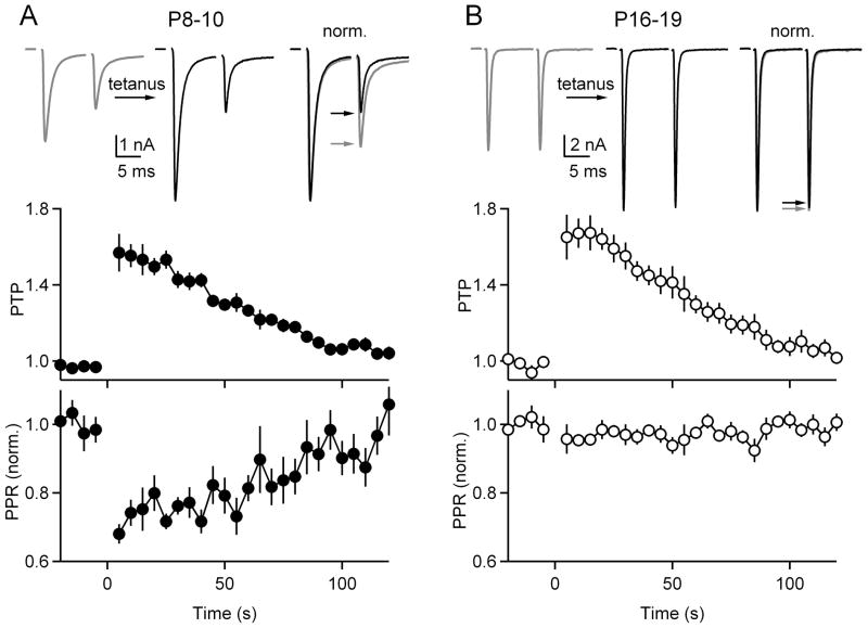
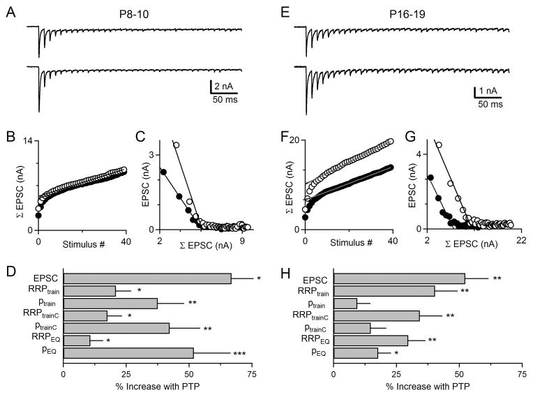
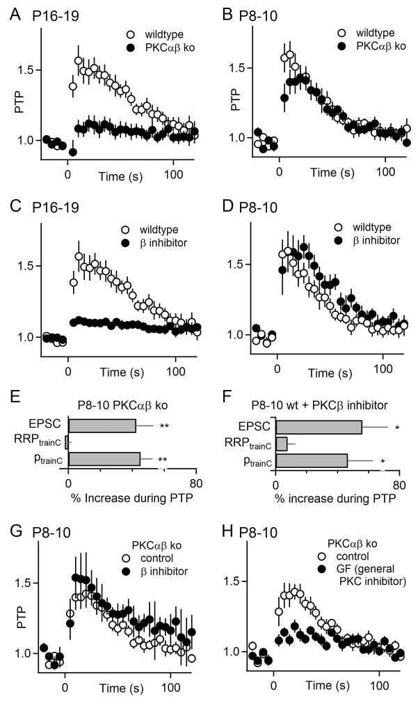
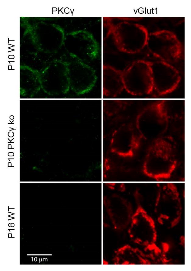
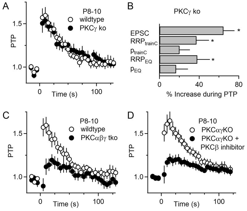
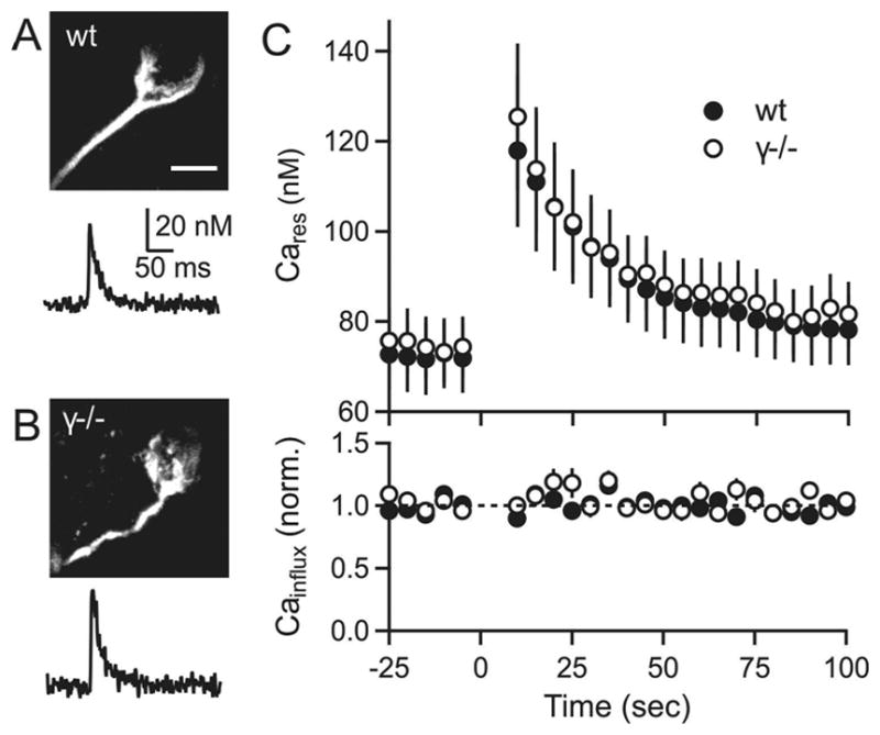
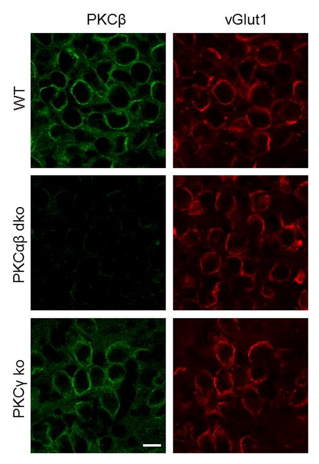
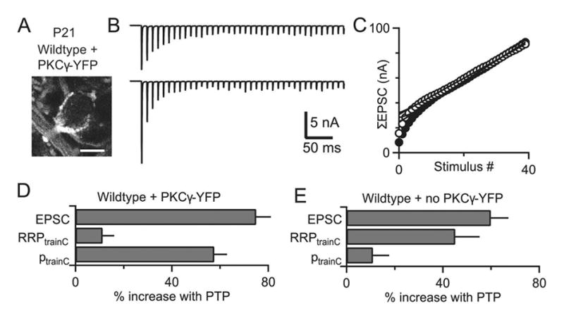
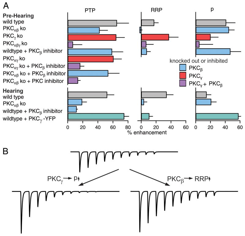
Posttetanic potentiation (PTP) is a widely observed form of short-term plasticity lasting for tens of seconds after high-frequency stimulation. Here we show that although protein kinase C (PKC) mediates PTP at the calyx of Held synapse in the auditory brainstem before and after hearing onset, PTP is produced primarily by an increased probability of release (p) before hearing onset, and by an increased readily releasable pool of vesicles (RRP) thereafter. We find that these mechanistic differences, which have distinct functional consequences, reflect unexpected differential actions of closely related calcium-dependent PKC isoforms. Prior to hearing onset, when PKCγ and PKCβ are both present, PKCγ mediates PTP by increasing p and partially suppressing PKCβ actions. After hearing onset, PKCγ is absent and PKCβ produces PTP by increasing RRP. In hearing animals, virally expressed PKCγ overrides PKCβ to produce PTP by increasing p. Thus, two similar PKC isoforms mediate PTP in distinctly different ways.


Figures

Figure 1
Figure 1. PTP is accompanied by a large decrease in paired-pulse plasticity before the onset of hearing but not after hearing onset
The calyx of Held was stimulated every 5 seconds with a pair of pulses separated by 10 ms, PTP was induced at t=0 using a 4 s, 100 Hz train and stimulation with pairs of pulses resumed. Results are shown for P8-10 animals (A) and P16-19 animals (B). Representative traces show the average baseline paired-pulse EPSC (A, B, top left, gray), the potentiated response during the peak of PTP (A, B, top middle, black), and traces normalized to the first EPSCs (A, B, top right). The EPSC amplitudes (A, B, middle) and the PPR are plotted as a function of time (A, B, bottom). Error bars are SEM.
Figure 2
Figure 2. Assessing the contributions of p and RRP to PTP before (A–D) and after (E–H) hearing onset
Mechanisms of PTP were examined using trains in the presence of kynurenate and CTZ to prevent receptor saturation and desensitization. (A, E) Synaptic currents evoked by the first 40 stimuli of 4s, 100 Hz train (top) and by a 40 pulse 100 Hz train (bottom) 10 s after tetanic stimulation (at the peak of PTP) are shown. The change in pool size and p were determined using the cumulative EPSC method, the corrected EPSC method (B, F) and the EQ method (C,G). (D, H) Summary of the changes in synaptic strength, RRP and p determined using the three different quantification methods. Paired t-tests were used to compare the changes in EPSC, RRPtrain, ptrain, RRPtrainC, ptrainC, RRPEQ, and pEQ during PTP to baseline, as indicated (*p<0.05, **p<0.01, ***p<0.001). Error bars are SEM.
Figure 3
Figure 3. Age-dependent differences in the roles of calcium-dependent PKC isoforms in PTP
PTP as a function of time for P16-19 animals (A, C) and P8-10 animals (B, D, G, H). (A, B) PTP from wildtype and PKCαβ dko animals are compared. (C, D) The effects of the PKCβ inhibitor (Calbiochem 539654) on PTP in wildtype animals are shown for the corresponding age groups. (E, F) Summary plots of RRP and p contributions to PTP in pre-hearing animals. Experiments were performed in the presence of CTZ and kynurenate as shown in Supplementary Figure S2. (G, H) The effects of the PKCβ inhibitor (G) and a broad spectrum PKC inhibitor (GF109203X) (H) on PTP in P8-10 PKCαβ dko animals are shown. *p<0.05, **p<0.01. Error bars are SEM.
Figure 4
Figure 4. Immunohistochemical localization of PKCγ at the calyx of Held in animals before and after hearing onset
Brain slices containing the MNTB region from wildtype and PKCγ ko animals prior to and after hearing onset were co-labeled with antibodies to PKCγ (green) and an antibody to the presynaptic marker vGlut1 (red). Representative images are shown for P10 and P18 slices from wildtype (WT) and PKCγ ko animals.
Figure 5
Figure 5. PKCγ mediates PTP in pre-hearing animals at the calyx of Held synapse
(A) PTP in P8-10 wildtype and PKCγ ko animals. (B) Summary of the contributions of RRP and p to synaptic enhancement in P8-10 PKCγ ko animals. Paired t-tests were used to compare the changes in EPSC, RRPtrainC, ptrainC during PTP to baseline, as indicated (*p<0.05). (C) PTP in P8-10 wildtype and PKCαβγ triple ko animals. (D) PTP in PKCαγ ko animals in the absence and the presence of a PKCβ inhibitor (250 nM). Error bars are SEM.
Figure 6
Figure 6. Tetanic stimulation produced similar presynaptic residual calcium signals in wildtype and PKCγ ko animals in pre-hearing animals
(A) top: two-photon image of a calyx from a wildtype animal filled with Alexa-594 dextran and calcium-green dextran. bottom: Calcium transient evoked by a single stimulus for a wildtype calyx. (B) Same as A, but for a PKCγ ko animal. (C) Plots of residual calcium (Cares, top) and calcium influx (bottom) in slices from wildtype (filled symbols) and PKCγ ko (open symbols) animals. Tetanic stimulation (4 s, 100 Hz) was at time t=0. Scale bar for (A, B) is 10 μm. Error bars are SEM.
Figure 7
Figure 7. PKCβ is present at the calyx of Held prior to hearing onset
Brain slices from P8-10 animals containing the MNTB region were co-labeled with antibodies to PKCβ (green) and an antibody to the presynaptic marker vGlut1 (red). Representative images are shown for P10 slices from wildtype (WT), PKCαβ ko, and PKCγ ko animals. Scale bar is 10 μm.
Figure 8
Figure 8. Viral expression of PKCγ in the calyx of Held of hearing animals alters the mechanism of PTP
An AAV expressing PKCγ-YFP was injected in the ventral cochlear nucleus at P4. Synaptic properties were examined at postnatal day 19–22 and calyces of Held expressing PKCγ-YFP were identified. Mechanisms of PTP were examined using trains in the presence of kynurenate and CTZ to prevent receptor saturation and desensitization. (A) Representative fluorescence image from a P21 wildtype calyx showing PKCγ-YFP fluorescence. (B) Synaptic currents evoked by the first 40 stimuli of 4s, 100 Hz train (top) and by a 40 pulse, 100 Hz train (bottom) 10 s after tetanic stimulation (at the peak of PTP) are shown. (C) Cumulative EPSCs for the initial train (closed circles) and the second train (open circles) are plotted against stimulus number. (D) Summary of the contributions of RRP and p to PTP for synapses expressing PKCγ-YFP. (E) Summary of the contributions of RRP and p to PTP for synapses not expressing PKCγ-YFP from the same animals as in D. Scale bar for (A) is 10 μm.
Figure 9
Figure 9. Summary and schematic showing the effects of different PKC isoforms on PTP before and after hearing onset
(A) Summary bar graphs are shown for the magnitude of PTP, and the changes in RRP and p that occur following tetanic stimulation in wildtype and knockout mice, and in the presence of pharmacological inhibitors. Bar graphs are color-coded to indicate whether PKCβ, PKCγ or both isoforms were knocked out or pharmacologically inhibited. Green bars indicate summaries of experiments in which PKCβ-YFP was expressed in wildtype animals. (B) A schematic illustration of the differential ways PKCβ and PKCγ contribute to PTP and the functional consequences of their response to a stimulus train.

Similar articles

Cited by

References

    1. Abbott LF, Regehr WG. Synaptic computation. Nature. 2004;431:796–803. - PubMed
    1. Abeliovich A, Chen C, Goda Y, Silva AJ, Stevens CF, Tonegawa S. Modified hippocampal long-term potentiation in PKC gamma-mutant mice. Cell. 1993;75:1253–1262. - PubMed
    1. Alle H, Jonas P, Geiger JR. PTP and LTP at a hippocampal mossy fiber-interneuron synapse. Proc Natl Acad Sci U S A. 2001;98:14708–14713. - PMC - PubMed
    1. Bao JX, Kandel ER, Hawkins RD. Involvement of pre- and postsynaptic mechanisms in posttetanic potentiation at Aplysia synapses. Science. 1997;275:969–973. - PubMed
    1. Barclay JW, Craig TJ, Fisher RJ, Ciufo LF, Evans GJ, Morgan A, Burgoyne RD. Phosphorylation of Munc18 by protein kinase C regulates the kinetics of exocytosis. J Biol Chem. 2003;278:10538–10545. - PubMed

Publication types

MeSH terms