. 2014 May 2;9(5):e96565.
doi: 10.1371/journal.pone.0096565. eCollection 2014.

Pancreatic ductal adenocarcinoma contains an effector and regulatory immune cell infiltrate that is altered by multimodal neoadjuvant treatment

Affiliations

Pancreatic ductal adenocarcinoma contains an effector and regulatory immune cell infiltrate that is altered by multimodal neoadjuvant treatment

Kendall C Shibuya et al. PLoS One. .

Abstract

Objective: The immune response to pancreatic ductal adenocarcinoma (PDA) may play a role in defining its uniquely aggressive biology; therefore, we sought to clearly define the adaptive immune infiltrate in PDA.

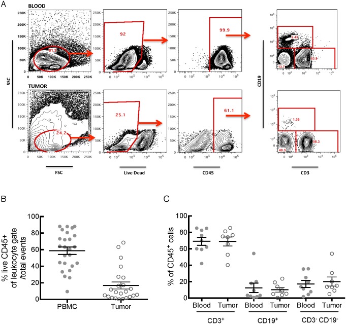
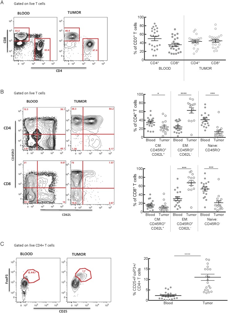
Design: We used immunohistochemistry and flow cytometry to characterize the immune infiltrate in human PDA and compared our findings to the patients' peripheral blood.

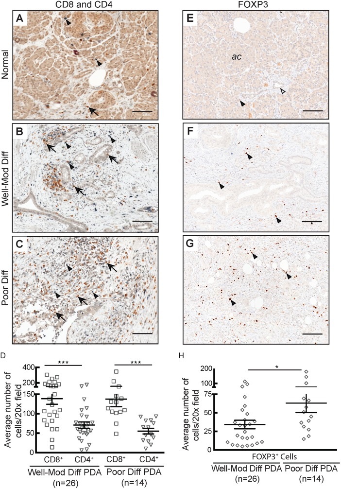
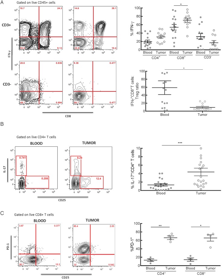
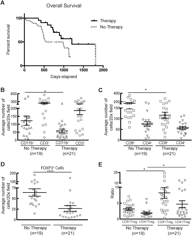
Results: In contrast to the myeloid cell predominant infiltrate seen in murine models, T cells comprised the majority of the hematopoietic cell component of the tumor stroma in human PDA. Most intratumoral CD8+ T cells exhibited an antigen-experienced effector memory cell phenotype and were capable of producing IFN-γ. CD4+ regulatory T cells (Treg) and IL-17 producing T helper cells were significantly more prevalent in tumor than in blood. Consistent with the association with reduced survival in previous studies, we observed higher frequencies of both myeloid cells and Treg in poorly differentiated tumors. The majority of intratumoral T cells expressed the co-inhibitory receptor programmed death-1 (PD-1), suggesting one potential mechanism through which PDA may evade antitumor immunity. Successful multimodal neoadjuvant therapy altered the immunoregulatory balance and was associated with reduced infiltration of both myeloid cells and Treg.

Conclusion: Our data show that human PDA contains a complex mixture of inflammatory and regulatory immune cells, and that neoadjuvant therapy attenuates the infiltration of intratumoral cells associated with immunosuppression and worsened survival.


Conflict of interest statement

Competing Interests: The authors have declared that no competing interests exist.

Figures

Figure 1
Figure 1. T cells infiltrate PDA regardless of GM-CSF expression.
A-C) IHC of normal adjacent pancreas, well-differentiated PDA, and poorly differentiated PDA showing dual staining of T cells (CD3+ in brown, arrows) and myeloid cells (CD11b+in purple, arrowheads). Open arrowhead indicates a normal duct. Photomicrographs taken with a 20x objective. D) Quantification of CD3+ and CD11b+ cells in PDA (p = 0.0009). E) Correlation between density of CD3+ and CD11b+ cells in each tumor (r = 0.51; p = 0.0008). F-H) Representative photomicrographs (40x) showing GM-CSF expression in normal pancreas and PDA. I) Distribution of myeloid cells and T cells stratified based upon chromogen intensity for GM-CSF. At least five 40x fields were analyzed per specimen. Scale bars = 25 µm.
Figure 2
Figure 2. T cells dominate the intratumoral immune cell infiltrate.
A) Representative flow cytometry plots illustrating the typical gating strategy used to identify the viable immune cells in blood and tumor. B) The proportion of live CD45+ cells was calculated for each specimen. C) CD3+ T cells consistently comprised the majority of the viable immune cells in blood and tumor, while CD19+ B cells and non-T or B cells comprise smaller proportions.
Figure 3
Figure 3. The majority of intratumoral T cells are antigen-experienced memory cells or regulatory cells.
A) Both CD4+ and CD8+ T cells infiltrate PDA. B) Representative plots and summed data of central memory (CM), effector memory (EM), and naïve CD4+ and CD8+ T cell populations in the blood and tumor among live CD3+ T cells. Tumors contained significantly more CD4+ (p<0.0001) and CD8+ (p = 0.0001) EM cells than did the blood, while containing fewer CD4+ CM (p = 0.01), CD4+ naïve (p = 0.0003) and CD8+ naïve (p = 0.0007) T cells. C) After gating on live CD3+ CD4+ T cells, we used CD25 and FOXP3 to identify putative regulatory T cells (p<0.0001).
Figure 4
Figure 4. Histologic localization of cellular subtypes confirms the flow cytometric analysis of the immune infiltrate in PDA.
A–C) Representative sections of normal pancreas and PDA showing multicolor IHC for CD8+ (brown, arrows) and CD4+ (purple, arrowheads) cells. D) Quantification of CD8+ (p = 0.0001) and CD4+ (p = 0.0005) cells. E–G) Representative sections of normal pancreas and PDA stained for FOXP3. Arrowheads indicate FOXP3+ cells, open arrowhead demonstrates a normal duct. H) Quantification of FOXP3+ cells (p = 0.02). At least five 20x fields were analyzed per specimen per cell type. Scale bars = 25 µm.
Figure 5
Figure 5. T cells in the tumor microenvironment retain functional capacity to activate inflammation despite the presence of multiple potential regulatory mechanisms.
A) Single-cell tumor suspensions and PBMC isolated from matched blood samples were incubated with PMA/ionomycin for 4 hours prior to staining for multiparameter flow cytometry. Staining for intracellular IFN-γ was positive in a significantly higher proportion of CD8+ T cells in tumor than in blood (p = 0.03), and there was a similar trend among CD4+ T cells (p = 0.05). A small proportion of CD3 cells thought to represent NK cells produced IFN-γ. The ratio of IFN-γ producing CD8+ T cells to Treg in blood and tumor is shown (p = 0.02). B) Th17 cells were identified by gating on live IL-17+CD4+ T cells. (p = 0.0008). C) Plot showed expression of PD-1 by both CD4+ and CD8+ unstimulated T cells (p = 0.003 and 0.02, respectively).
Figure 6
Figure 6. Multimodal neoadjuvant therapy alters the regulatory cell makeup of the tumor microenvironment.
A) Kaplan-Meier survival of patients in this study. There was a trend towards longer survival among patients undergoing neoadjuvant therapy (p = 0.07). B) Quantification of CD11b+ (p = 0.04) and CD3+cells in neoadjuvant therapy vs. untreated tumors. C) CD4+ cells were unchanged, however CD8+ cells were less frequent after neoadjuvant therapy (p = 0.04). D) Quantification of FOXP3+ cells in treated and untreated tumors (p = 0.002). E) Analysis of ratio of CD8+ to FOXP3+ T cells (p = 0.01) and CD4+ to FOXP3+ T cells (p = 0.01) in treated and untreated tumors.

References

    1. Siegel R, Naishadham D, Jemal A (2012) Cancer statistics, 2012. CA: A Cancer Journal for Clinicians 62: 10–29 10.3322/caac.20138 - DOI - PubMed
    1. Ino Y, Yamazaki-Itoh R, Shimada K, Iwasaki M, Kosuge T, et al. (2013) Immune cell infiltration as an indicator of the immune microenvironment of pancreatic cancer. Br J Cancer 108: 914–923 10.1038/bjc.2013.32 - DOI - PMC - PubMed
    1. Katz SC, Bamboat ZM, Maker AV, Shia J, Pillarisetty VG, et al. (2013) Regulatory T cell infiltration predicts outcome following resection of colorectal cancer liver metastases. Ann Surg Oncol 20: 946–955 10.1245/s1043401226689 - DOI - PMC - PubMed
    1. Milne K, Köbel M, Kalloger SE, Barnes RO, Gao D, et al. (2009) Systematic Analysis of Immune Infiltrates in High-Grade Serous Ovarian Cancer Reveals CD20, FoxP3 and TIA-1 as Positive Prognostic Factors. PLoS ONE 4: e6412 10.1371/journal.pone.0006412.t003 - DOI - PMC - PubMed
    1. Hingorani SR, Wang L, Multani AS, Combs C, Deramaudt TB, et al. (2005) Trp53R172H and KrasG12D cooperate to promote chromosomal instability and widely metastatic pancreatic ductal adenocarcinoma in mice. Cancer Cell 7: 469–483 10.1016/j.ccr.2005.04.023 - DOI - PubMed

Publication types

MeSH terms

Substances