. 2014 May 22;7(4):1310-9.
doi: 10.1016/j.celrep.2014.03.071. Epub 2014 May 1.

Patterns of somatically acquired amplifications and deletions in apparently normal tissues of ovarian cancer patients

Affiliations

Patterns of somatically acquired amplifications and deletions in apparently normal tissues of ovarian cancer patients

Leila Aghili et al. Cell Rep. .

Abstract

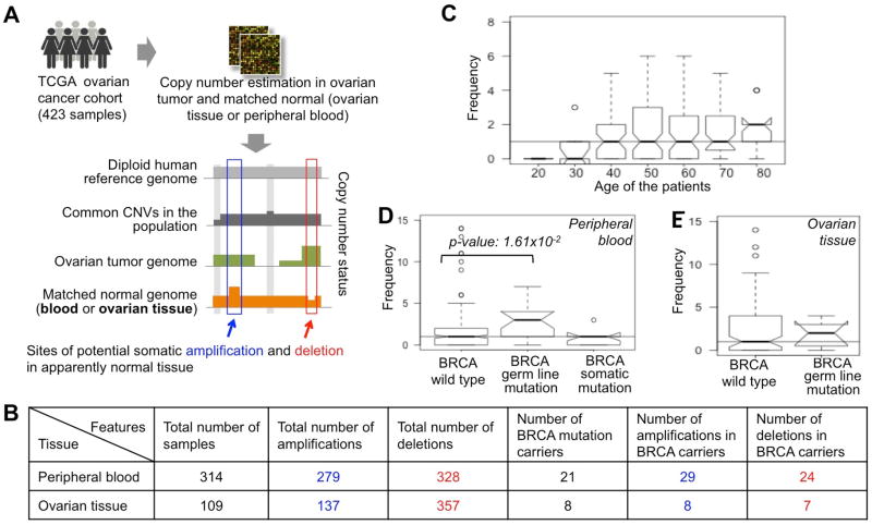
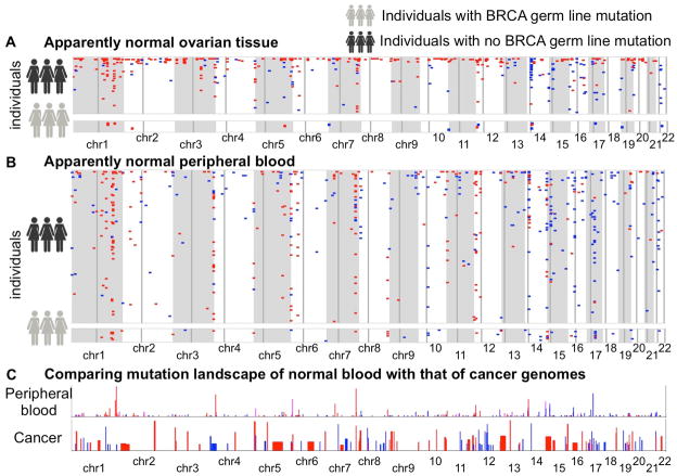
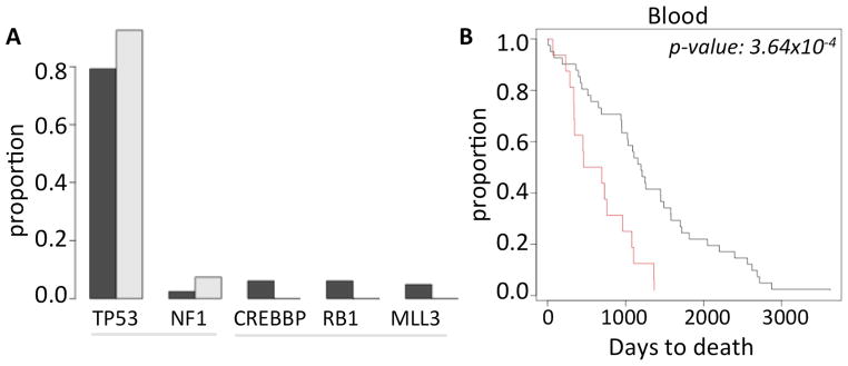
Little is understood about the occurrence of somatic genomic alterations in normal tissues and their significance in the context of disease. Here, we identified potential somatic copy number alterations (pSCNAs) in apparently normal ovarian tissue and peripheral blood of 423 ovarian cancer patients. There were, on average, two to four pSCNAs per sample detectable at a tissue-level resolution, although some individuals had orders of magnitude more. Accordingly, we estimated the lower bound of the rate of pSCNAs per cell division. Older individuals and BRCA mutation carriers had more pSCNAs than others. pSCNAs significantly overlapped with Alu and G-quadruplexes, and the affected genes were enriched for signaling and regulation. Some of the amplification/deletion hotspots in pan-cancer genomes were hot spots of pSCNAs in normal tissues as well, suggesting that those regions might be inherently unstable. Prevalence of pSCNA in peripheral blood predicted survival, implying that mutations in normal tissues might have consequences for cancer patients.


Figures

Figure 1
Figure 1
(A) The pipeline for detecting of somatic amplifications and deletions in apparently normal peripheral blood and ovarian cancer tissue of the TCGA ovarian cancer patients. (B) Summary statistics of somatic amplifications and deletions in peripheral blood and ovarian cancer tissue in the cohort. The BRCA mutation carriers are shown categorically. (C-E) The number of somatic genomic alterations (amplifications and deletions) per (C) peripheral blood sample, grouped according to the age of the individuals, (D) peripheral blood sample, grouped according to BRCA mutation status, and (E) ovarian tissue sample, grouped according to BRCA mutation status. The horizontal line shows the median value across all the samples in respective panel.
Figure 2
Figure 2
Genome-wide mutational landscape showing pSCNAnorm ( amplifications: blue, deletions: red) in apparently normal (A) ovarian cancer tissue and (B) peripheral blood of the ovarian cancer patients. Each row represents an individual. Chromosomes are indicated below. Faint vertical lines in each chromosome indicate centromere. Only the individuals with at least one detectable pSCNAnorm in the cohort are shown. The individuals with germ line BRCA1 or BRCA2 mutations are shown separately. (C) Comparing the mutation landscape of apparently normal peripheral blood with that of 26 different cancer types combined, as analyzed by Beroukhim et al. Nature. 2010. The balance of blue and red shades indicates the proportion of amplifications and deletions. Heights of the bars indicate prevalence of such events in the genome.
Figure 3
Figure 3
(A) Frequency of the common cancer gene mutations in ovarian cancer patients who had no pSCNAbl (black) and those who have ≥4 pSCNAbl (grey). (B) Kaplan Meier curve showing difference in the survival patterns between the ovarian cancer patients who have no pSCNAbl (black) and those who have ≥4 pSCNAbl (red). The difference was statistically significant ( log rank test, p-value 3.64×10⁻⁴).

Similar articles

Cited by

References

    1. Abyzov A, Mariani J, Palejev D, Zhang Y, Haney MS, Tomasini L, Ferrandino AF, Rosenberg Belmaker LA, Szekely A, Wilson M, et al. Somatic copy number mosaicism in human skin revealed by induced pluripotent stem cells. Nature. 2012;492:438–442. - PMC - PubMed
    1. Aoyama K, Nagata M, Oshima K, Matsuda T, Aoki N. Molecular cloning and characterization of a novel dual specificity phosphatase, LMW-DSP2, that lacks the cdc25 homology domain. The Journal of biological chemistry. 2001;276:27575–27583. - PubMed
    1. Araten DJ, Golde DW, Zhang RH, Thaler HT, Gargiulo L, Notaro R, Luzzatto L. A quantitative measurement of the human somatic mutation rate. Cancer research. 2005;65:8111–8117. - PubMed
    1. Bannert N, Vollhardt K, Asomuddinov B, Haag M, Konig H, Norley S, Kurth R. PDZ Domain-mediated interaction of interleukin-16 precursor proteins with myosin phosphatase targeting subunits. The Journal of biological chemistry. 2003;278:42190–42199. - PubMed
    1. Barlow JH, Faryabi RB, Callen E, Wong N, Malhowski A, Chen HT, Gutierrez-Cruz G, Sun HW, McKinnon P, Wright G, et al. Identification of early replicating fragile sites that contribute to genome instability. Cell. 2013;152:620–632. - PMC - PubMed

Publication types